
Specialty Medical genetics DiseasesDB 28840 | ICD-9-CM 277.87 MeSH D028361 | |
 | ||
Similar Friedreich's ataxia, Pearson syndrome, Wolfram syndrome | ||
Mitochondrial disease is a group of disorders caused by dysfunctional mitochondria, the organelles that generate energy for the cell. Mitochondria are found in every cell of the human body except red blood cells, and convert the energy of food molecules into the ATP that powers most cell functions.
Contents
- Signs and symptoms
- Causes
- Examples
- Mechanisms
- Detection
- Treatments
- Epidemiology
- History
- Notable cases
- References
Mitochondrial diseases are sometimes (about 15% of the time) caused by mutations in the mitochondrial DNA that affect mitochondrial function. Other causes of mitochondrial disease are mutations in genes of the nuclear DNA, whose gene products are imported into the mitochondria (mitochondrial proteins) as well as acquired mitochondrial conditions. Mitochondrial diseases take on unique characteristics both because of the way the diseases are often inherited and because mitochondria are so critical to cell function. The subclass of these diseases that have neuromuscular disease symptoms are often called a mitochondrial myopathy.
Signs and symptoms
Symptoms include poor growth, loss of muscle coordination, muscle weakness, visual problems, hearing problems, learning disabilities, heart disease, liver disease, kidney disease, gastrointestinal disorders, respiratory disorders, neurological problems, autonomic dysfunction and dementia.
The body, and each mutation, is modulated by other genome variants; the mutation that in one individual may cause liver disease might in another person cause a brain disorder. The severity of the specific defect may also be great or small. Some minor defects cause only "exercise intolerance", with no serious illness or disability. Defects often affect the operation of the mitochondria and multiple tissues more severely, leading to multi-system diseases.
As a rule, mitochondrial diseases are worse when the defective mitochondria are present in the muscles, cerebrum, or nerves, because these cells use more energy than most other cells in the body.
Although mitochondrial diseases vary greatly in presentation from person to person, several major clinical categories of these conditions have been defined, based on the most common phenotypic features, symptoms, and signs associated with the particular mutations that tend to cause them.
An outstanding question and area of research is whether ATP depletion or reactive oxygen species are in fact responsible for the observed phenotypic consequences.
Causes
Mitochondrial disorders may be caused by mutations, acquired or inherited, in mitochondrial DNA (mtDNA) or in nuclear genes that code for mitochondrial components. They may also be the result of acquired mitochondrial dysfunction due to adverse effects of drugs, infections, or other environmental causes (see MeSH).
Nuclear DNA has two copies per cell (except for sperm and egg cells), one copy being inherited from the father and the other from the mother. Mitochondrial DNA, however, is strictly inherited from the mother and each mitochondrial organelle typically contains between 2 to 10 mtDNA copies. During cell division the existing mitochondria segregate randomly between the two new cells, and then those mitochondria make more copies, normally reaching 500 mitochondria per cell. As mtDNA is copied when mitochondria proliferate, they can accumulate random mutations, a phenomenon called heteroplasmy. If only a few of the mtDNA copies inherited from the mother are defective, mitochondrial division may cause most of the defective copies to end up in just one of the new mitochondria (for more detailed inheritance patterns, see Human mitochondrial genetics). Mitochondrial disease may become clinically apparent once the number of affected mitochondria reaches a certain level; this phenomenon is called "threshold expression".
Mitochondrial DNA mutations occur frequently, due to the lack of the error checking capability that nuclear DNA has (see Mutation rate). This means that mitochondrial DNA disorders may occur spontaneously and relatively often. Defects in enzymes that control mitochondrial DNA replication (all of which are encoded for by genes in the nuclear DNA) may also cause mitochondrial DNA mutations.
Most mitochondrial function and biogenesis is controlled by nuclear DNA. Human mitochondrial DNA encodes only 13 proteins of the respiratory chain, while most of the estimated 1,500 proteins and components targeted to mitochondria are nuclear-encoded. Defects in nuclear-encoded mitochondrial genes are associated with hundreds of clinical disease phenotypes including anemia, dementia, hypertension, lymphoma, retinopathy, seizures, and neurodevelopmental disorders.
A study by Yale University researchers published in the Feb 12, 2004 issue of the New England Journal of Medicine explores the role of mitochondria in insulin resistance among the offspring of patients with type 2 diabetes. Other studies have shown that the mechanism may involve the interruption of the mitochondrial signaling process in body cells (intramyocellular lipids). A study conducted at the Pennington Biomedical Research Center in Baton Rouge, LA (Diabetes 54, 2005 1926-33) showed that this in turn partially disables the genes that produce mitochondria.
Examples
Examples of mitochondrial diseases include:
Nota bene: Conditions such as Friedreich's ataxia can affect the mitochondria, but are not associated with mitochondrial proteins.
Mechanisms
The effective overall energy unit for the available body energy is referred to as the daily glycogen generation capacity, and is used to compare the mitochondrial output of healthy individuals to that of afflicted or chronically glycogen-depleted individuals. This value is slow to change in a given individual, as it takes between 18 and 24 months to complete a full cycle.
The glycogen generation capacity is entirely dependent on, and determined by, the operating levels of the mitochondria in all of the cells of the human body; however, the relation between the energy generated by the mitochondria and the glycogen capacity is very loose and is mediated by many biochemical pathways. The energy output of full healthy mitochondrial function can be predicted exactly by a complicated theoretical argument, but this argument is not straightforward, as most energy is consumed by the brain and is not easily measurable.
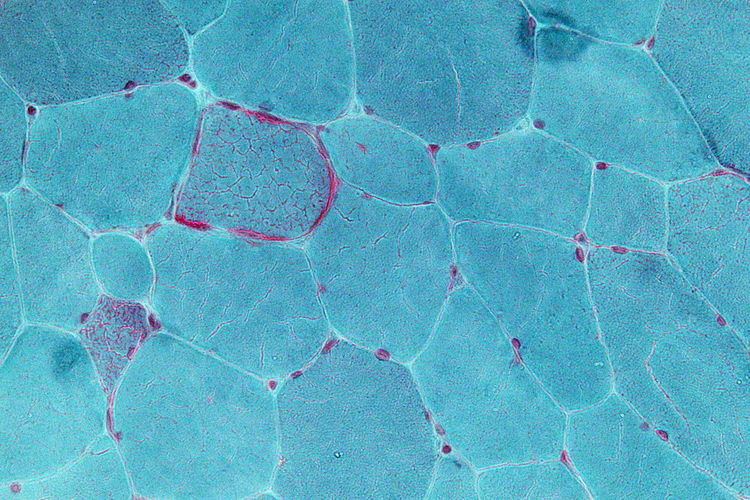
Detection
Mitochondrial diseases are usually detected by analysing muscle samples, where the presence of these organelles is higher. The most common tests for the detection of these diseases are:
- Southern blot to detect big deletions or duplications
- PCR and specific mutaltation analysis
- Sequencing
Treatments
Although research is ongoing, treatment options are currently limited; vitamins are frequently prescribed, though the evidence for their effectiveness is limited. Membrane penetrating antioxidants, such as the mitochondria-targeted antioxidant MitoQ (mitoquinol mesylate) have the most important role in improving mitochondrial dysfunction. Pyruvate has been proposed recently as a treatment option. N acetylcysteine reverses many models of mitochondrial dysfunction.
Spindle transfer, where the nuclear DNA is transferred to another healthy egg cell leaving the defective mitochondrial DNA behind, is a potential treatment procedure that has been successfully carried out on monkeys. Using a similar pronuclear transfer technique, researchers at Newcastle University led by Douglass Turnbull successfully transplanted healthy DNA in human eggs from women with mitochondrial disease into the eggs of women donors who were unaffected. In such cases, ethical questions have been raised regarding biological motherhood, since the child receives genes and gene regulatory molecules from two different women. Using genetic engineering in attempts to produce babies free of mitochondrial disease is controversial in some circles and raises important ethical issues. A male baby was born in Mexico in 2016 from a mother with Leigh syndrome using spindle transfer.
In September 2012 a public consultation was launched in the UK to explore the ethical issues involved. Human genetic engineering was used on a small scale to allow infertile women with genetic defects in their mitochondria to have children. In June 2013, the United Kingdom government agreed to develop legislation that would legalize the 'three-person IVF' procedure as a treatment to fix or eliminate mitochondrial diseases that are passed on from mother to child. The procedure could be offered from 29 October 2015 once regulations had been established. Embryonic mitochondrial transplant and protofection have been proposed as a possible treatment for inherited mitochondrial disease, and allotopic expression of mitochondrial proteins as a radical treatment for mtDNA mutation load.
Currently, human clinical trials are underway at GenSight Biologics (ClinicalTrials.gov # NCT02064569) and the University of Miami (ClinicalTrials.gov # NCT02161380) to examine the safety and efficacy of mitochondrial gene therapy in Leber's hereditary optic neuropathy.
Epidemiology
About 1 in 4,000 children in the United States will develop mitochondrial disease by the age of 10 years. Up to 4,000 children per year in the US are born with a type of mitochondrial disease. Because mitochondrial disorders contain many variations and subsets, some particular mitochondrial disorders are very rare.
The average number of births per year among women at risk for transmitting mtDNA disease is estimated to approximately 150 in the United Kingdom and 800 in the United States.
History
The first pathogenic mutation in mitochondrial DNA was identified in 1988, from that time to 2016, around 275 other disease-causing mutations were identified.
Notable cases
Notable people who suffered from mitochondrial disease include: