
Latin lemniscus medialis NeuroLex ID Medial lemniscus FMA 83675 | NeuroNames ancil-736 Dorlands/Elsevier l_06/12483115 | |
 | ||
TA A14.1.04.111A14.1.08.672 | ||
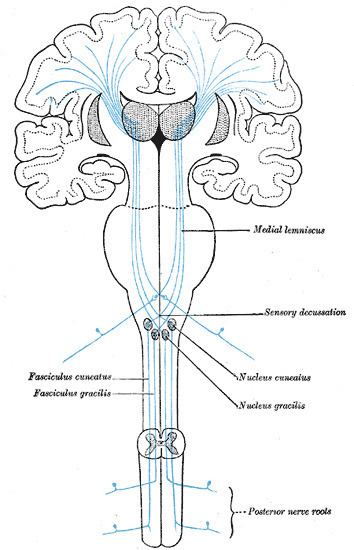
The medial lemniscus, also known as Reil's band or Reil's ribbon, is a large ascending bundle of heavily myelinated axons that decussate in the brainstem, specifically in the medulla oblongata. The medial lemniscus is formed by the crossings of the internal arcuate fibers. The internal arcuate fibers are composed of axons of nucleus gracilis and nucleus cuneatus. The axons of the nucleus gracilis and nucleus cuneatus in the medial lemniscus have cell bodies that lie contralaterally.
Contents
The medial lemniscus is part of the posterior column–medial lemniscus pathway, which ascends from the skin to the thalamus, which is important for somatosensation from the skin and joints, therefore, lesion of the medial lemnisci causes an impairment of vibratory and touch-pressure sense.
Etymology
Lemniscus means "ribbon", so named because the medial lemniscus "spirals" or "turns" as it ascends.
Path
After neurons carrying proprioceptive or touch information synapse at the gracile and cuneate nuclei, axons from secondary neurons decussate at the level of the medulla and travel up the brainstem as the medial lemniscus on the contralateral (opposite) side. It is part of the posterior column-medial lemniscus pathway, which transmits touch, vibration sense, as well as the pathway for proprioception.
The medial lemniscus carries axons from most of the body and synapses in the ventral posterolateral nucleus of the thalamus, at the level of the mamillary bodies. Sensory axons transmitting information from the head and neck via the trigeminal nerve synapse at the ventral posteromedial nucleus of the thalamus.