
 | ||
Masson's trichrome is a three-colour staining protocol used in histology. The recipes evolved from Claude L. Pierre Masson's (1880–1959) original formulation have different specific applications, but all are suited for distinguishing cells from surrounding connective tissue.
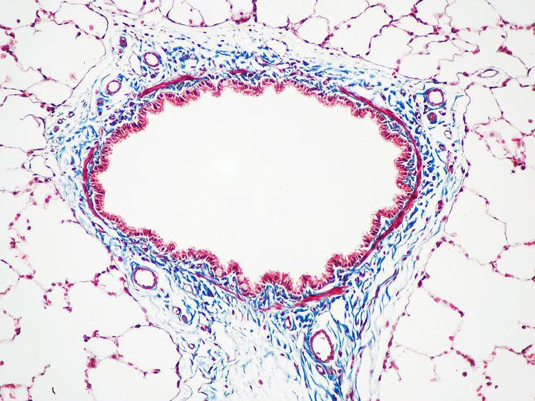
Most recipes produce red keratin and muscle fibers, blue or green collagen and bone, light red or pink cytoplasm, and dark brown to black cell nuclei.
The trichrome is applied by immersion of the fixated sample into Weigert's iron hematoxylin, and then three different solutions, labeled A, B, and C:
Standard applications: Masson’s trichrome staining is widely used to study muscular pathologies (muscular dystrophy), cardiac pathologies (infarct), hepatic pathologies (cirrhosis) or kidney pathologies (glomerular fibrosis). It can also be used to detect and analyze routinely tumors on hepatic and kidney biopsies.
Variants
A common variant is Lillie's trichrome. It is often erroneously called Masson's trichrome. It differs in the dyes used, their concentrations, and the immersion times.
Another common variant is the Masson trichrome & Verhoeff stain, which combines the Masson trichrome stain and Verhoeff stain. This combination is useful for the examination of blood vessels; the Verhoeff stain highlights elastin (black) and allows one to easily differentiate small arteries (which typically have at least two elastic laminae) and veins (which have one elastic lamina).