
 | ||
Single probe application mass spectrometry imaging and single cell analysis
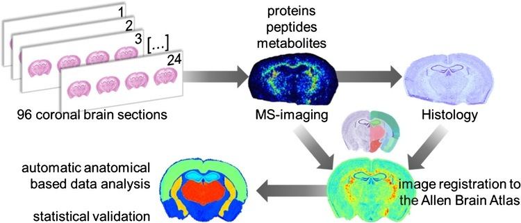
Mass spectrometry imaging (MSI) (also known as imaging mass spectrometry) is a technique used in mass spectrometry to visualize the spatial distribution of chemical compositions e.g. compounds, biomarkers, metabolites, peptides or proteins by their molecular masses. Although widely used traditional methodologies like radiochemistry and immunohistochemistry achieve the same goal as MSI, they are limited in their abilities to analyze multiple samples at once, and can prove to be lacking if researchers do not have prior knowledge of the samples being studied. Emerging technologies in the field of MSI are MALDI imaging and secondary ion mass spectrometry imaging (SIMS imaging).
Contents
- Single probe application mass spectrometry imaging and single cell analysis
- Coming to a hospital near you mass spectrometry imaging
- SIMS imaging
- MALDI imaging
- Ambient ionization methods
- Standard data format for mass spectrometry imaging datasets
- Software
- References
Coming to a hospital near you mass spectrometry imaging
SIMS imaging
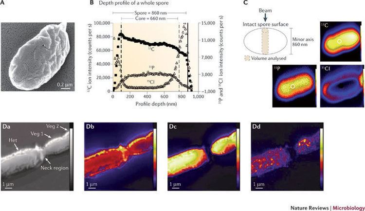
Secondary ion mass spectrometry (SIMS) is used to analyze solid surfaces and thin films by sputtering the surface with a focused primary ion beam and collecting and analyzing ejected secondary ions. There are many different sources for a primary ion beam. However, the primary ion beam must contain ions that are at the higher end of the energy scale. Some common sources are: Cs+, O2+, O, Ar+ and Ga+. SIMS imaging is performed in a manner similar to electron microscopy; the primary ion beam is emitted across the sample while secondary mass spectra are recorded. SIMS proves to be advantageous in providing increased resolution for visualizing spatial distribution over smaller mass ranges. SIMS is widely regarded as one of the most sensitive forms of mass spectrometry as it can detect elements as small as 10−6-10−9.
Multiplexed ion beam imaging (MIBI) is a SIMS method that uses metal isotope labeled antibodies to label compounds in biological samples.
Developments within SIMS: Some chemical modifications have been made within SIMS to increase the efficiency of the process. There are currently two separate techniques being used to help increase the overall efficiency by increasing the sensitivity of SIMS measurements: matrix-enhanced SIMS (ME-SIMS) - This has the same sample preparation as MALDI does as this simulates the chemical ionization properties of MALDI. ME-SIMS does not sample nearly as much material. However, if the analyte being tested has a low mass value then it can produce a similar looking spectra to that of a MALDI spectra. ME-SIMS has been so effective that it has been able to detect low mass chemicals at sub cellular levels that was not possible prior to the development of the ME-SIMS technique. The second technique being used is called sample metallization (Meta-SIMS) - This is the process of gold or silver addition to the sample. This forms a layer of gold or silver around the sample and it is normally no more than 1-3 nm thick. Using this technique has resulted in an increase of sensitivity for larger mass samples. The addition of the metallic layer also allows for the conversion of insulating samples to conducting samples, thus charge compensation within SIMS experiments is no longer required.
MALDI imaging
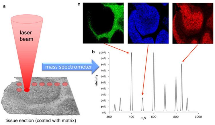
Matrix-assisted laser desorption ionization can be used as a mass spectrometry imaging technique for relatively large molecules. It has recently been proven that the most effective type of matrice to use is an ionic matrice for MALDI imaging of tissue. In this version of the technique the sample, typically a thin tissue section, is moved in two dimensions while the mass spectrum is recorded. Although MALDI has the benefit of being able to record spatial distribution of larger molecules, it comes at the cost of worse resolution than the SIMS technique. The power source normally used in MALDI experiments for ionization to occur is either an Nd:YAG (355 nm) or N2 (337 nm) laser.
Pharmacodynamics and toxicodynamics in tissue have been studied by MALDI imaging.
Ambient ionization methods
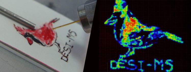
Ambient ionization methods such as desorption electrospray ionization (DESI) and matrix-assisted laser desorption electrospray ionization (MALDESI) have also been employed for MSI.
Standard data format for mass spectrometry imaging datasets
As an extension of the mzML standard format developed by HUPO for mass spectrometry datasets, the Computis project developed the imzML standard format for mass spectrometry imaging datasets.
Software
There are large amounts of free software packages available for visualization and mining of imaging mass spectrometry data. Converters from Thermo Fisher format, Analyze format, GRD format and Bruker format to imzML format were developed by the Computis project. Some software modules are also available for viewing mass spectrometry images in imzML format: Biomap] (Novartis, free), Datacube Explorer (AMOLF, free), EasyMSI (CEA), Mirion (JLU), MSiReader (NCSU, free), SpectralAnalysis (https://github.com/AlanRace/SpectralAnalysis).
For processing .imzML files with the free statistical and graphics language R, a collection of R scripts are available, which also permits parallel-processing of large files on a local computer, a remote cluster or on the Amazon cloud (http://www.bioprocess.org/msi.r).
Another free statistical package for processing imzML and Analyze 7.5 data in R exists, Cardinal (http://cardinalmsi.org/).
In addition the following commercial software suites for MALDI imaging are available: MALDIVision (Premier Biosoft viewing software, http://www.premierbiosoft.com/maldi-tissue-imaging/index.html), Quantinetix, and SCiLS Lab(statistical add-on program to Bruker's FlexImaging, http://scils.de/).