
 | ||
Origin Spinous processes of vertebrae T7-T12, thoracolumbar fascia, iliac crest, inferior 3 or 4 ribs and inferior angle of scapula Nerve Thoracodorsal nerve(C6,C7,C8) Actions Adducts, extends and internally rotates the arm when the insertion is moved towards the origin. When observing the muscle action of the origin towards the insertion, the lats are a very powerful rotator of the trunk. Antagonist Deltoid and trapezius muscle | ||
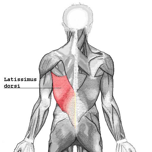
The latissimus dorsi (/ˌləˈtɪsᵻməs ˈdɔːrsaɪ/) (plural: latissimi dorsi), meaning 'broadest [muscle] of the back' (Latin latus meaning 'broad', latissimus meaning 'broadest' and dorsum meaning the back), is the larger, flat, dorso-lateral muscle on the trunk, posterior to the arm, and partly covered by the trapezius on its median dorsal region. Latissimi dorsi are commonly known as "lats", especially among bodybuilders.
Contents
The latissimus dorsi is responsible for extension, adduction, transverse extension also known as horizontal abduction, flexion from an extended position, and (medial) internal rotation of the shoulder joint. It also has a synergistic role in extension and lateral flexion of the lumbar spine.
Due to bypassing the scapulothoracic joints and attaching directly to the spine, the actions the latissimi dorsi have on moving the arms can also influence the movement of the scapulae, such as their downward rotation during a pull up.
Variations
The number of dorsal vertebrae to which it is attached varies from four to eight; the number of costal attachments varies; muscle fibers may or may not reach the crest of the ilium.
A muscular slip, the axillary arch, varying from 7 to 10 cm in length, and from 5 to 15 mm in breadth, occasionally springs from the upper edge of the latissimus dorsi about the middle of the posterior fold of the axilla, and crosses the axilla in front of the axillary vessels and nerves, to join the under surface of the tendon of the pectoralis major, the coracobrachialis, or the fascia over the biceps brachii. This axillary arch crosses the axillary artery, just above the spot usually selected for the application of a ligature, and may mislead a surgeon. It is present in about 7% of the population and may be easily recognized by the transverse direction of its fibers. Guy et al. extensively described this muscular variant using MRI data and positively correlated its presence with symptoms of neurological impingement.
A fibrous slip usually passes from the upper border of the tendon of the Latissimus dorsi, near its insertion, to the long head of the triceps brachii. This is occasionally muscular, and is the representative of the dorsoepitrochlearis brachii of apes. This muscular form is found in ~5% of humans and is sometimes termed the latissimocondyloideus.[2]
The latissimus dorsi crosses the inferior angle of the scapula. A study found that, of 100 cadavers dissected:
Triangles
Innervation
The latissimus dorsi is supplied by the sixth, seventh, and eighth cervical nerves through the thoracodorsal (long scapular) nerve. Electromyography suggests that it consists of six groups of muscle fibres that can be independently coordinated by the central nervous system.
Function
The latissimus dorsi is responsible for extension, adduction, transverse extension also known as horizontal abduction, flexion from an extended position, and (medial) internal rotation of the shoulder joint. It also has a synergistic role in extension (posterior fibers) and lateral flexion (anterior fibers) of the lumbar spine, and assists as a muscle of both forced expiration (anterior fibers) and an accessory muscle of inspiration (posterior fibers).
Most latissimus dorsi exercises concurrently recruit the teres major, posterior fibres of the deltoid, long head of the triceps brachii, among numerous other stabilizing muscles. Compound exercises for the 'lats' typically involve elbow flexion and tend to recruit the biceps brachii, brachialis, and brachioradialis for this function. Depending on the line of pull, the trapezius muscles can be recruited as well; horizontal pulling motions such as rows recruit both latissimus dorsi and trapezius heavily.
Training
The power/size/strength of this muscle can be trained with a variety of different exercises. Some of these include:
Clinical relevance
Tight latissimus dorsi has been shown to be one cause of chronic shoulder pain and chronic back pain. Because the latissimus dorsi connects the spine to the humerus, tightness in this muscle can manifest as either sub-optimal glenohumeral joint (shoulder) function which leads to chronic pain or tendinitis in the tendinous fasciae connecting the latissimus dorsi to the thoracic and lumbar spine.
The latissimus dorsi is a potential source of muscle for breast reconstruction surgery after mastectomy (e.g. Mannu flap) or to correct pectoral hypoplastic defects such as Poland's syndrome. An absent or hypoplastic latissimus dorsi can be one of the associated symptoms of Poland's syndrome.
Cardiac support
For heart patients with low cardiac output and who are not candidates for cardiac transplantation, a procedure called cardiomyoplasty may support the failing heart. This procedure involves wrapping the latissimus dorsi muscles around the heart and electrostimulating them in synchrony with ventricular systole.