
Dorlands/Elsevier l_09/12491634 FMA 58283 | TA A04.4.02.007 | |
 | ||
Latin ligamentum arcuatum laterale | ||
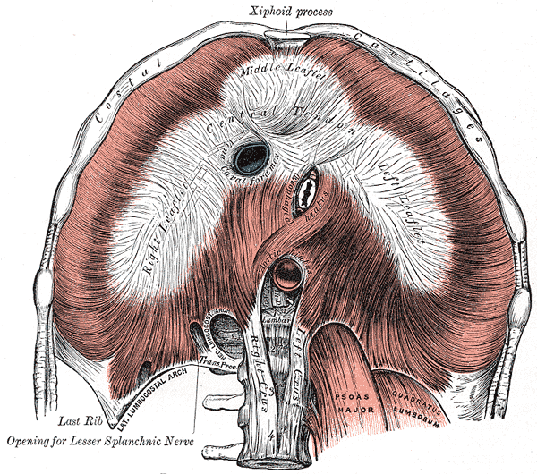
The lateral arcuate ligament (also lateral lumbocostal arch and external arcuate ligament) is a ligament under the diaphragm that arches across the upper part of the quadratus lumborum muscle. It is traversed by the subcostal nerve, artery and vein.
Contents
Structure
The lateral arcuate ligament runs from the front of the transverse process of the first lumbar vertebra, and, laterally, to the tip and lower margin of the twelfth rib. It forms an arch over the quadratus lumborum muscle.
Variations
Although the lateral arcuate ligament is commonly described in anatomy textbooks as attaching at the first lumbar vertebra (L1) other instances have been found in cadaver studies with attachments at either the second (L2) or third (L3) lumbar vertebra.
In some people (~5%) inferolateral extensions of the lateral arcuate ligaments present as thickened nodular areas adjacent to the lateral diaphragmatic surface which can be visualized with computed tomography (CT) scans.
History
The lateral arcuate ligaments were described by Galen, as early as AD 177, from animal dissections performed as part of his Rome lectures, collected in De Anatomicus Administrationibus.