Kalpana Kalpana (Editor)

Jugular foramen

Updated on
Edit
Like
Comment
Share on FacebookTweet on TwitterShare on LinkedInShare on Reddit
Latin
  
Foramen jugulare

FMA
  
56432

TA
  
A02.1.00.054

Jugular foramen

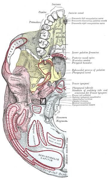
The jugular foramen is a large foramen (aperture) in the base of the skull. It is located behind the carotid canal and is formed in front by the petrous portion of the temporal bone, and behind by the occipital bone; it is generally larger on the right than on the left side.

Contents

Contents

Cranial nerves IX, X, and XI and the internal jugular vein pass through the jugular foramen.

The jugular foramen may be subdivided into three compartments, each with their own contents.

  • The anterior compartment transmits the inferior petrosal sinus and glossopharyngeal nerve (CN IX)
  • The intermediate transmits the vagus and accessory nerves (aka cranial nerves number X, and XI respectively).
  • The posterior transmits the sigmoid sinus (becoming the internal jugular vein) and some meningeal branches from the occipital and ascending pharyngeal arteries.
  • An alternative imaging based subclassification exists, delineated by the jugular spine which is a bony ridge partially separating the jugular foramen into two parts:

  • The smaller, anteromedial, "pars nervosa" compartment contains CN IX, (tympanic nerve, a branch of CN IX), and receives the venous return from inferior petrosal sinus.
  • The larger, posterolateral, "pars vascularis" compartment contains CN X, CN XI, Arnold's nerve (or the auricular branch of CN X involved in the Arnold's reflex, where external auditory meatus stimulation causes cough), jugular bulb, and posterior meningeal branch of ascending pharyngeal artery.
  • Clinical significance

    Obstruction can result in "Vernet's syndrome".

    References

    Jugular foramen Wikipedia