
 | ||
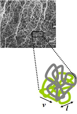
Intravoxel incoherent motion (IVIM) imaging is a concept and a method initially introduced and developed by Le Bihan et al. to quantitatively assess all the microscopic translational motions that could contribute to the signal acquired with diffusion MRI. In biological tissue, these motions essentially are molecular diffusion of water and microcirculation of blood in the capillary network (perfusion). The concept introduced by D. Le Bihan is that water flowing in randomly oriented capillaries (at the voxel level) mimics a random walk (“pseudo-diffusion” ) (Fig.1).
Contents
It is responsible for a signal attenuation in diffusion MRI, which depends on the velocity of the flowing blood and the vascular architecture. Similarly to molecular diffusion, the effect of pseudodiffusion on the signal attenuation depends on the b value. However, the rate of signal attenuation resulting from pseudodiffusion is typically an order of magnitude greater than molecular diffusion in tissues, so its relative contribution to the diffusion-weighted MRI signal becomes significant only at very low b values, allowing diffusion and perfusion effects to be separated.
Model
In the presence of the magnetic field gradient pulses of a diffusion MRI sequence, the MRI signal gets attenuated due to diffusion and perfusion effects. In a simple model, this signal attenuation, S/So, can be written as:
where
Assuming blood water flowing in the randomly oriented vasculature changes several times direction (at least 2) during the measurement time (model 1), one has for
where
where
If blood water flows without changing direction (either because flow is slow or measurement time is short) while capillary segments are randomly and isotropically oriented (model 2),
where
In both cases, the perfusion effect results in a curvature of the diffusion attenuation plot towards b=0 (Fig.2).
In a simple approach and under some approximations, the ADC calculated from 2 diffusion-weighted images acquired with b0=0 and b1, as ADC = ln(S(b0)/S (b1)), is:
where
Biexponential model:
Where
Kurtosis model:
where
Applications
IVIM MRI was initially introduced to evaluate perfusion and produce maps of brain perfusion, for brain activation studies (before the introduction of BOLD fMRI) and clinical applications (stroke, brain tumors). Recent work has proven the validity of the IVIM concept from fMRI, with an increase in the IVIM perfusion parameters in brain activated regions, and the potential of the approach to aid in our understanding of the different vascular contributions to the fMRI signal. IVIM MRI has also been used in the context of fMRI in a negative way.
A limitation of BOLD fMRI is its spatial resolution, as flow increase in somewhat large arteries or veins feed or drain large neuronal territories. By inserting “diffusion” gradient pulses in the MRI sequence (corresponding to low b-values), one may crush the contribution of the largest vessels (with high D* values associated with fast flow) in the BOLD signal and improve the spatial resolution of the activation maps. Several groups have relied on this trick, though not always considering referring to the IVIM concept. This IVIM concept has also been borrowed to improve other applications, for instance, arterial spin labeling (ASL) or to suppress signal from extracellular flowing fluid in perfused cell systems.
However, IVIM MRI has recently undergone a striking revival for applications not in the brain, but throughout the body as well. Following earlier encouraging results in the kidneys, or even the heart, IVIM MRI really took off for liver applications. For instance, Luciani et al. found that D* was significantly reduced in cirrhotic patients, which, according to the IVIM model, points out to reduce blood velocity (and flow). (Another theoretical, rather unlikely interpretation would be that capillary segments become longer or more straight in those patients with liver fibrosis). The perfusion fraction, f, which is linked to blood volume in the IVIM model, remained normal, confirming earlier results by Yamada et al. Though, blood volume is expected to be reduced in liver cirrhosis.
One has to keep in mind that IVIM imaging has a differential sensitivity to vessel types, according to the range of motion sensitization (b values) which are used. Signal from large vessels with rapid flow disappears quickly with very low b values, while smaller vessels with slower flow might still contribute to the IVIM signal acquired with b values larger than 200 s/mm². Many more applications are now under investigation, especially for imaging of patients suspected of cancer in the body (prostate, liver, kidney, pancreas, etc.) and human placenta. A key feature of IVIM diffusion MRI is that it does not involve contrast agents, and it may appear as an interesting alternative for perfusion MRI in some patients at risk for Nephrogenic Systemic Fibrosis (NSF).