
 | ||
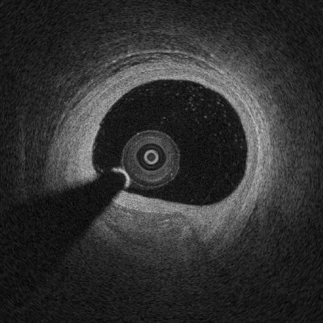
Intracoronary optical coherence tomography (OCT) (or, more generally, intravascular optical coherence tomography, IVOCT), is an endoscopic-based application of optical coherence tomography. Analogous to IVUS, intracoronary OCT uses a catheter to deliver and collect near infrared light (e.g., 1,300 nm) to create cross-sectional images images of artery lumen. Intracoronary OCT creates images at a resolution of approximately 15 micro-meters, an order of magnitude improved resolution with respect to IVUS and x-ray coronary angiogram.
Contents
Medical uses
Approximately 100,000 intracoronary optical coherence tomography procedures are performed every year and its adoption is rapidly growing at a rate of ~20% every year. Evidence showed that intracoronary OCT can be used to optimize percutaneous coronary intervention (PCI) to treat myocardial infarction and that OCT imaging influence physician decision in >50% of the cases.
Assessment of artery lumen morphology is the cornerstone of intravascular imaging criteria to evaluate disease severity and guide intervention. The high-resolution of OCT imaging allows to assess with high accuracy vessel lumen area, wall microstructure, intracoronary stent apposition and expansion. OCT has an improved ability with respect to IVUS to penetrate and delineate calcium in the vessel wall that makes it well suited to guide complex interventional strategies in vessels with superficial calcification. OCT has the capability of visualize coronary plaque erosion and fibrotic caps overlying atheromas.
Safety
Safety of intravascular imaging, including intracoronary OCT and intravascular ultrasound, has been investigated by several studies. Recent clinical trials reported a very low rate of self-limiting, minor complications on over 3,000 patients where in all cases no harm or prologation of hospital stay was observed. Intracoronary optical coherence tomography was demonstrates to be safe among heterogeneous groups of patients presenting varying clinical setting.
Methods
State-of-the-art intracoronary optical coherence tomography typically uses a swept-source laser to acquire OCT images at high-speed of approximately 80,000 kHz (A-scan lines per second) to complete acquisition of a 3D OCT volume of a coronary segments in a few-seconds.