Suvarna Garge (Editor)

Interventricular septum

Updated on
Edit
Like
Comment
Share on FacebookTweet on TwitterShare on LinkedInShare on Reddit
MeSH
  
A07.541.459

TA
  
A12.1.00.013

Dorlands /Elsevier
  
s_08/12730379

FMA
  
7133

Interventricular septum

Artery
  
anterior interventricular branch of left coronary artery and Posterior interventricular artery

Latin
  
s. interventriculare cordis

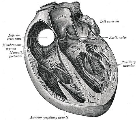
The interventricular septum (IVS), (or ventricular septum, or during development septum inferius), is the stout wall separating the lower chambers (the ventricles) of the heart from one another.

Contents

The ventricular septum is directed obliquely backward to the right, and curved with the convexity toward the right ventricle; its margins correspond with the anterior and posterior longitudinal sulci.

Portions

  • The greater portion of it is thick and muscular and constitutes the muscular interventricular septum.
  • Its upper and posterior part, which separates the aortic vestibule from the lower part of the right atrium and upper part of the right ventricle, is thin and fibrous, and is termed the membranous ventricular septum (septum membranaceum)..
  • Development

  • The muscular part of the interventricular septum derives from the bulboventricular flange which is developed due to differential growth of primitive ventricle and bulbous cordis. Membranous part has a neural crest origin which connects the upper free margin of the bulboventricular flange and anterior and posterior endocardial cushions of atrio ventricular canal. It also gets attached to lower border of spiral septum or the aortico pulmonary septum.
  • Disorders

  • Acyanotic heart defect
  • Ventricular septal defect (VSD), a hole in the interventricular septum
  • References

    Interventricular septum Wikipedia