
ICD-O M8825/1 | ||
 | ||
Inflammatory myofibroblastic tumour is a lesional pattern of inflammatory pseudotumour, as plasma cell granuloma. It is abbreviated IMT.
Contents
Symptoms
The symptoms depend on the specific location of the tumour, which can be anywhere in the body.
Diagnosis
Inflammatory myofibroblastic tumours are diagnosed based on their appearance under the microscope, by pathologists. Medical imaging findings are non-specific.
Pathology
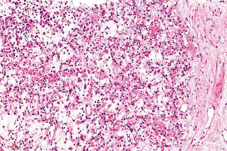
Inflammatory myofibroblastic tumours are characterized by a mix of inflammatory cells, e.g. plasma cells, lymphocytes and eosinophils, and bland spindle cells without nuclear atypia. These tumours may have necrosis, hemorrhage, focal calcification and mitotic activity.
The histologic differential diagnosis includes:
Approximately half of IMTs have a rearrangement of the ALK gene.