
 | ||
Hyperpolarization is a change in a cell's membrane potential that makes it more negative. It is the opposite of a depolarization. It inhibits action potentials by increasing the stimulus required to move the membrane potential to the action potential threshold.
Contents
Hyperpolarization is often caused by efflux of K+ (a cation) through K+ channels, or influx of Cl– (an anion) through Cl– channels. On the other hand, influx of cations, e.g. Na+ through Na+ channels or Ca2+ through Ca2+ channels, inhibits hyperpolarization. If a cell has Na+ or Ca2+ currents at rest, then inhibition of those currents will also result in a hyperpolarization. This voltage-gated ion channel response is how the hyperpolarization state is achieved. In neurons, the cell enters a state of hyperpolarization immediately following the generation of an action potential. While hyperpolarized, the neuron is in a refractory period that lasts roughly 2 milliseconds, during which the neuron is unable to generate subsequent action potentials. Sodium-potassium ATPases redistribute K+ and Na+ ions until the membrane potential is back to its resting potential of around –70 millivolts, at which point the neuron is once again ready to transmit another action potential.
Voltage-gated ion channels and hyperpolarization
Voltage gated ion channels respond to changes in the membrane potential, generating the action potential as well as hyperpolarization. These channels work by selecting an ion based on electrostatic attraction or repulsion allowing the ion to bind to the channel. This releases water molecules attached to the ion, and the ion passes through the pore.
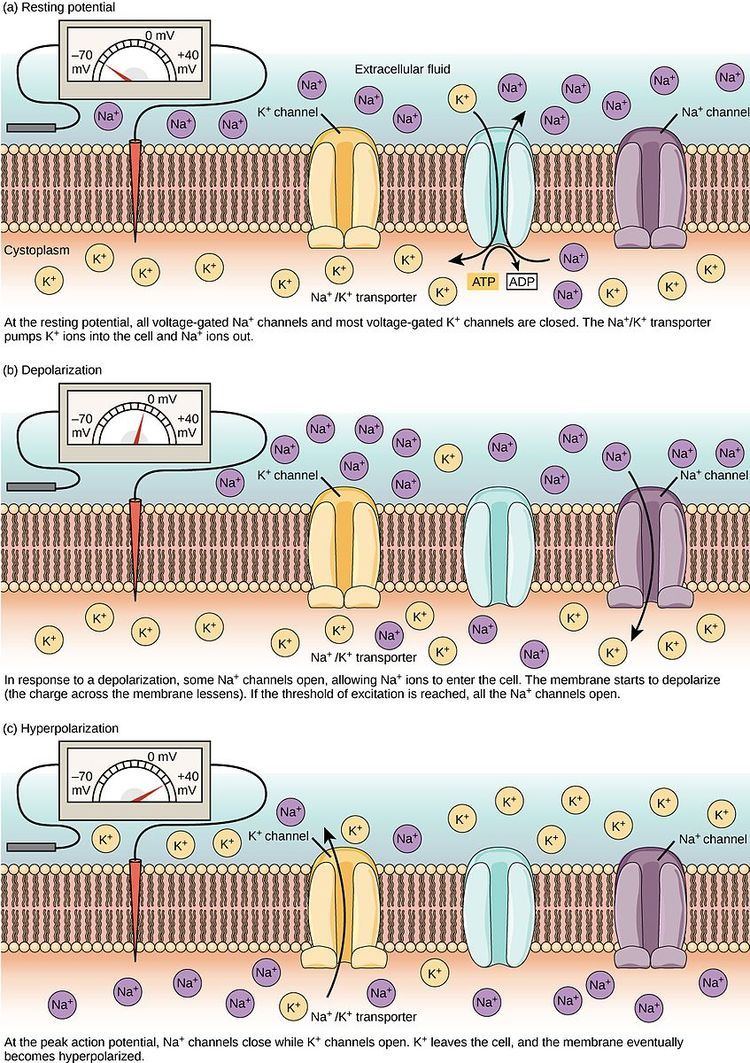
At resting potential, the voltage gated sodium and potassium channels are both closed, but as the cell membrane begins to become depolarized, the voltage gated sodium channels begin to open and the neuron begins to depolarize even more, creating a current feedback loop known as the Hodgkin cycle. However, since potassium ions can leak out of the cell to counteract the sodium channel inflow, if the original depolarization event was not large enough the neuron will not generate an action potential. If however a large number of sodium channels are opened, the neuron becomes approximately ten times more permeable to sodium than to potassium, leading quickly to depolarization of the cell to a peak near +40 mV. At this voltage the sodium channels begin to inactivate and voltage gated potassium channels begin to open. This combination of closed sodium channels (no more positive charge flowing into the neuron) and open potassium channels (positively charged potassium ions leaving the cell) leads the neuron to become hyperpolarized. The neuron continues to repolarize until the cell reaches approximately –75 mV, which is the reversal potential of potassium ions. This results in an afterhyperpolarization, which leads to a refractory period, during which the neuron is incapable of triggering another action potential due to the sodium channels' inability to open. At this point the voltage gated potassium channels close and the resting permeability of the membrane to sodium and potassium allows the neuron to return to its resting potential of –70 mV.
HCN channels are activated by hyperpolarization.
Experimental technique
Hyperpolarization is a change in membrane potential, neuroscientists measure it using a technique known as patch clamping. Using this method they are able to record ion currents passing through individual channels. This is done using a glass micropipette, also called a patch pipette, with a 1 micrometer diameter. There is a small patch that contains a few ion channels and the rest is sealed off, making this the point of entry for the current. Using an amplifier and a voltage clamp, which is an electronic feedback circuit, allows the experimenter to maintain the membrane potential at a fixed point and the voltage clamp then measures tiny changes in current flow. The membrane currents giving rise to hyperpolarization are either an increase in outward current or a decrease in inward current.
Examples
- During the afterhyperpolarization period after an action potential, the membrane potential is more negative than when the cell is at the resting potential. In the figure to the right, this undershoot occurs at approximately 3 to 4 milliseconds (ms) on the time scale. The afterhyperpolarization is the time when the membrane potential is hyperpolarized relative to the resting potential.
- During the rising phase of an action potential, the membrane potential changes from negative to positive, a depolarization. In the figure, the rising phase is from approximately 1 to 2 ms on the graph. During the rising phase, once the membrane potential becomes positive, the membrane potential continues to depolarize (overshoot) until the peak of the action potential is reached at about +40 millivolts (mV). After the peak of the action potential, a hyperpolarization repolarizes the membrane potential to its resting value, first by making it less positive, until 0 mV is reached, and then by continuing to make it more negative. This repolarization occurs in the figure from approximately 2 to 3 ms on the time scale.