
Synonyms haematoma ICD-10 M79.81 DiseasesDB 5487 | ICD-9-CM 729.92 MeSH D006406 | |
 | ||
A hematoma is a localized collection of blood outside the blood vessels, due to either disease or trauma including injury or surgery and may involve blood continuing to seep from broken capillaries. A hematoma is initially in liquid form spread among the tissues including in sacs between tissues where it may coagulate and solidify before blood is reabsorbed into blood vessels. An ecchymosis is a hematoma of the skin larger than 10mm.
Contents
They may occur among/within many areas such as fat, skin and other organs, connective tissues, bone, joints and muscle.
A collection of blood (or even a hemorrhage) may be aggravated by an anticoagulant, "blood thinner". Blood seepage and collection of blood may occur if heparin is given via an intramuscular route; to avoid this, heparin must be given intravenously or subcutaneously.
It is not to be confused with hemangioma, which is an abnormal buildup/growth of blood vessels in the skin or internal organs.
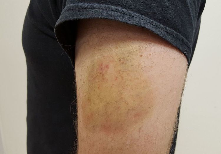
Signs and symptoms
Some hematomas are visible under the surface of the skin (commonly called bruises) or possibly felt as masses/lumps. Lumps may be caused by the limitation of the blood to a sac, subcutaneous or intramuscular tissue space isolated by fascial planes. This is a key anatomical feature that helps prevent injuries from causing massive blood loss. In most cases the hematoma such as a sac of blood eventually dissolves; however, in some cases they may continue to grow such as due to blood seepage or show no change. If the sac of blood does not disappear, then it may need to be surgically cleaned out/repaired.
The slow process of reabsorption of hematomas can allow the broken down blood cells and hemoglobin pigment to move in the connective tissue. For example, a patient who injures the base of his thumb might cause a hematoma, which will slowly move all through the finger within a week. Gravity is the main determinant of this process.
Hematomas on articulations can reduce mobility of a member and present roughly the same symptoms as a fracture.
In most cases, movement and exercise of the affected muscle is the best way to introduce the collection back into the blood stream.
A mis-diagnosis of a hematoma in the vertebra can sometimes occur; this is correctly called a hemangioma (buildup of cells) or a benign tumor.
Types
Degrees
Etymology
The word "haematoma" came into use around 1850. The word derives from the Greek roots "heme-" (blood) and -oma, from soma, meaning body = a body of blood. Another etymological derivation would be from "haemat-" and "-oma" = "-ing", thus simply "bleeding".