
Species Human Entrez 3098 | Human Mouse Ensembl ENSG00000156515 | |
 | ||
Aliases HK1, HK1-ta, HK1-tb, HK1-tc, HKD, HKI, HMSNR, HXK1, hexokinase 1, HK, hexokinase External IDs MGI: 96103 HomoloGene: 100530 GeneCards: HK1 | ||
Hexokinase-1 (HK1) is an enzyme that in humans is encoded by the HK1 gene on chromosome 10. Hexokinases phosphorylate glucose to produce glucose-6-phosphate (G6P), the first step in most glucose metabolism pathways. This gene encodes a ubiquitous form of hexokinase which localizes to the outer membrane of mitochondria. Mutations in this gene have been associated with hemolytic anemia due to hexokinase deficiency. Alternative splicing of this gene results in five transcript variants which encode different isoforms, some of which are tissue-specific. Each isoform has a distinct N-terminus; the remainder of the protein is identical among all the isoforms. A sixth transcript variant has been described, but due to the presence of several stop codons, it is not thought to encode a protein. [provided by RefSeq, Apr 2009]
Contents
Structure
HK1 is one of four highly homologous hexokinase isoforms in mammalian cells.
Gene
The HK1 gene spans approximately 131 kb and consists of 25 exons. Alternative splicing of its 5’ exons produces different transcripts in different cell types: exons 1-5 and exon 8 (exons T1-6) are testis-specific exons; exon 6, located approximately 15 kb downstream of the testis-specific exons, is the erythroid-specific exon (exon R); and exon 7, located approximately 2.85 kb downstream of exon R, is the first 5’ exon for the ubiquitously expressed HK1 isoform. Moreover, exon 7 encodes the porin-binding domain (PBD) conserved in mammalian HK1 genes. Meanwhile, the remaining 17 exons are shared among all HK1 isoforms.
In addition to exon R, a stretch of the proximal promoter that contains a GATA element, an SP1 site, CCAAT, and an Ets-binding motif is necessary for expression of HK-R in erythroid cells.
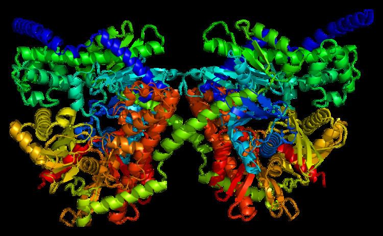
Protein
This gene encodes a 100 kDa homodimer with a regulatory N-terminal domain (1-475), catalytic C-terminal domain (residues 476-917), and an alpha-helix connecting its two subunits. Both terminal domains are composed of a large subdomain and a small subdomain. The flexible region of the C-terminal large subdomain (residues 766–810) can adopt various positions and is proposed to interact with the base of ATP. Moreover, glucose and G6P bind in close proximity at the N- and C-terminal domains and stabilize a common conformational state of the C-terminal domain. According to one model, G6P acts as an allosteric inhibitor which binds the N-terminal domain to stabilize its closed conformation, which then stabilizes a conformation of the C-terminal flexible subdomain that blocks ATP. A second model posits that G6P acts as an active inhibitor that stabilizes the closed conformation and competes with ATP for the C-terminal binding site. Results from several studies suggest that the C-terminal is capable of both catalytic and regulatory action. Meanwhile, the hydrophobic N-terminal lacks enzymatic activity by itself but contains the G6P regulatory site and the PBD, which is responsible for the protein’s stability and binding to the outer mitochondrial membrane (OMM).
Function
As one of two mitochondrial isoforms of hexokinase and a member of the sugar kinase family, HK1 catalyzes the rate-limiting and first obligatory step of glucose metabolism, which is the ATP-dependent phosphorylation of glucose to G6P. Physiological levels of G6P can regulate this process by inhibiting HK1 as negative feedback, though inorganic phosphate (Pi) can relieve G6P inhibition. However, unlike HK2 and HK3, HK1 itself is not directly regulated by Pi, which better suits its ubiquitous catabolic role. By phosphorylating glucose, HK1 effectively prevents glucose from leaving the cell and, thus, commits glucose to energy metabolism. Moreover, its localization and attachment to the OMM promotes the coupling of glycolysis to mitochondrial oxidative phosphorylation, which greatly enhances ATP production by direct recycling of mitochondrial ATP/ADP to meet the cell’s energy demands. Specifically, OMM-bound HK1 binds VDAC1 to trigger opening of the mitochondrial permeability transition pore and release mitochondrial ATP to further fuel the glycolytic process.
Another critical function for OMM-bound HK1 is cell survival and protection against oxidative damage. Activation of Akt kinase is mediated by HK1-VDAC1 coupling as part of the growth factor-mediated phosphatidyl inositol 3-kinase (PI3)/Akt cell survival intracellular signaling pathway, thus preventing cytochrome c release and subsequent apoptosis. In fact, there is evidence that VDAC binding by the anti-apoptotic HK1 and by the pro-apoptotic creatine kinase are mutually exclusive, indicating that the absence of HK1 allows creatine kinase to bind and open VDAC. Furthermore, HK1 has demonstrated anti-apoptotic activity by antagonizing Bcl-2 proteins located at the OMM, which then inhibits TNF-induced apoptosis.
In the prefrontal cortex, HK1 putatively forms a protein complex with EAAT2, Na+/K+ ATPase, and aconitase, which functions to remove glutamate from the perisynaptic space and maintain low basal levels in the synaptic cleft.
In particular, HK1 is the most ubiquitously expressed isoform out of the four hexokinases, and constitutively expressed expressed in most tissues, though it is majorly found in brain, kidney, and red blood cells (RBCs). Its high abundance in the retina, specifically the photoreceptor inner segment, outer plexiform layer, inner nuclear layer, inner plexiform layer, and ganglion cell layer, attests to its crucial metabolic purpose. It is also expressed in cells derived from hematopoietic stem cells, such as RBCs, leukocytes, and platelets, as well as from erythroid-progenitor cells. Of note, HK1 is the sole hexokinase isoform found in the cells and tissues which rely most heavily on glucose metabolism for their function, including brain, erythrocytes, platelets, leukocytes, and fibroblasts. In rats, it is also the predominant hexokinase in fetal tissues, likely due to their constitutive glucose utilization.
Clinical significance
Mutations in this gene are associated with type 4H of Charcot–Marie–Tooth disease, also known as Russe-type hereditary motor and sensory neuropathy (HMSNR). Due to the crucial role of HK1 in glycolysis, hexokinase deficiency has been identified as a cause of erythroenzymopathies associated with hereditary non-spherocytic hemolytic anemia (HNSHA). Likewise, HK1 deficiency has resulted in cerebral white matter injury, malformations, and psychomotor retardation, as well as latent diabetes mellitus and panmyelopathy. Meanwhile, HK1 is highly expressed in cancers, and its anti-apoptotic effects have been observed in highly glycolytic hepatoma cells.
Neurodegenerative disorders
HK1 may be causally linked to mood and psychotic disorders, including unipolar depression (UPD), bipolar disorder (BPD), and schizophrenia via both its roles in energy metabolism and cell survival. For instance, the accumulation of lactate in the brains of BPD and SCHZ patients potentially results from the decoupling of HK1 from the OMM, and by extension, glycolysis from mitochondrial oxidative, phosphorylation. In the case of SCHZ, decreasing HK1 attachment to the OMM in the parietal cortex resulted in decreased glutamate reuptake capacity and, thus, glutamate spillover from the synapses. The released glutamate activates extrasynaptic glutamate receptors, leading to altered structure and function of glutamate circuits, synaptic plasticity, frontal cortical dysfunction, and ultimately, the cognitive deficits characteristic of SCHZ. Similarly, Hk1 mitochondrial detachment has been associated with hypothyroidism, which involves abnormal brain development and increased risk for depression, while its attachment leads to neural growth. In Parkinson’s disease, HK1 detachment from VDAC via Parkin-mediated ubiquitylation and degradation disrupts the MPTP on depolarized mitochondria, consequently blocking mitochondrial localization of Parkin and halting glycolysis. Further research is required to determine the relative HK1 detachment needed in various cell types for different psychiatric disorders. This research can also contribute to developing therapies to target causes of the detachment, from gene mutations to interference by factors such as beta-amyloid peptide and insulin.
Retinitis pigmentosa
A heterozygous missense mutation in the HK1 gene (a change at position 847 from glutamate to lysine) has been linked to retinitis pigmentosa. Since this substitution mutation is located far from known functional sites and does not impair the enzyme’s glycolytic activity, it is likely that the mutation acts through another biological mechanism unique to the retina. Notably, studies in mouse retina reveal interactions between Hk1, the mitochondrial metallochaperone Cox11, and the chaperone protein Ranbp2, which serve to maintain normal metabolism and function in the retina. Thus, the mutation may disrupt these interactions and lead to retinal degradation. Alternatively, this mutation may act through the enzyme’s anti-apoptotic function, as disrupting the regulation of the hexokinase-mitochondria association by insulin receptors could trigger photoreceptor apoptosis and retinal degeneration. In this case, treatments that preserve the hexokinase–mitochondria association may serve as a potential therapeutic approach.
Interactions
HK1 is known to interact with:
Interactive pathway map
Click on genes, proteins and metabolites below to link to respective articles.