
EC number 2.7.10.1 Human Mouse | Species Human Entrez 2064 | |
 | ||
Aliases ERBB2, CD340, HER-2, HER-2/neu, HER2, MLN 19, NEU, NGL, TKR1, erb-b2 receptor tyrosine kinase 2 External IDs OMIM: 164870 MGI: 95410 HomoloGene: 3273 GeneCards: ERBB2 | ||
Receptor tyrosine-protein kinase erbB-2, also known as CD340 (cluster of differentiation 340), proto-oncogene Neu, Erbb2 (rodent), or ERBB2 (human). It is a protein that in humans is encoded by the ERBB2 gene, and it is also frequently called HER2 (from human epidermal growth factor receptor 2) or HER2/neu.
Contents
- Name
- Gene
- Function
- Signal transduction
- HER2 and cancer
- HER2 variationsmutations
- Drugs targeting HER2
- HER2 testing
- HER2 testing on tumour
- HER2 testing on serum
- HER2 interactions
- References
HER2 is a member of the human epidermal growth factor receptor (HER/EGFR/ERBB) family. Amplification or over-expression of this oncogene has been shown to play an important role in the development and progression of certain aggressive types of breast cancer. In recent years the protein has become an important biomarker and target of therapy for approximately 30% of breast cancer patients.
Name
HER2 is so named because it has a similar structure to human epidermal growth factor receptor, or HER1. Neu is so named because it was derived from a rodent glioblastoma cell line, a type of neural tumor. ErbB-2 was named for its similarity to ErbB (avian erythroblastosis oncogene B), the oncogene later found to code for EGFR. Molecular cloning of the gene showed that HER2, Neu, and ErbB-2 are all encoded by the same orthologs.
Gene
ERBB2, a known proto-oncogene, is located at the long arm of human chromosome 17 (17q12).
Function
The ErbB family consists of four plasma membrane-bound receptor tyrosine kinases. One of which is erbB-2, and the other members being epidermal growth factor receptor, erbB-3 (neuregulin-binding; lacks kinase domain), and erbB-4. All four contain an extracellular ligand binding domain, a transmembrane domain, and an intracellular domain that can interact with a multitude of signaling molecules and exhibit both ligand-dependent and ligand-independent activity. Notably, no ligands for HER2 have yet been identified. HER2 can heterodimerise with any of the other three receptors and is considered to be the preferred dimerisation partner of the other ErbB receptors.
Dimerisation results in the autophosphorylation of tyrosine residues within the cytoplasmic domain of the receptors and initiates a variety of signaling pathways.
Signal transduction
Signaling pathways activated by HER2 include:
In summary, signaling through the ErbB family of receptors promotes cell proliferation and opposes apoptosis, and therefore must be tightly regulated to prevent uncontrolled cell growth from occurring.
HER2 and cancer
Amplification, also known as the over-expression of the ERBB2 gene, occurs in approximately 15-30% of breast cancers. It is strongly associated with increased disease recurrence and a poor prognosis. Over-expression is also known to occur in ovarian, stomach, adenocarcinoma of the lung and aggressive forms of uterine cancer, such as uterine serous endometrial carcinoma, e.g. HER-2 is over-expressed in approximately 7-34% of patients with gastric cancer and in 30% of salivary duct carcinomas.
HER2 is co-localised, and, most of the time, co-amplified with the gene GRB7, which is a proto-oncogene associated with breast, testicular germ cell, gastric, and eosophageal tumours.
HER2 proteins have been shown to form clusters in cell membranes that may play a role in tumorigenesis.
Recent evidence has implicated HER2 signaling in resistance to the EGFR-targeted cancer drug cetuximab.
HER2 variations/mutations
Furthermore, diverse structural alterations have been identified that cause ligand-independent firing of this receptor, doing so in the absence of receptor over-expression. HER2 is found in a variety of tumours and some of these tumours carry point mutations in the sequence specifying the transmembrane domain of HER2. Substitution of a valine for a glutamic acid in the transmembrane domain can result in the constitutive dimerisation of this protein in the absence of a ligand.
HER2 mutations have been found in non-small-cell lung cancers (NSCLC) and can direct treatment.
Drugs targeting HER2
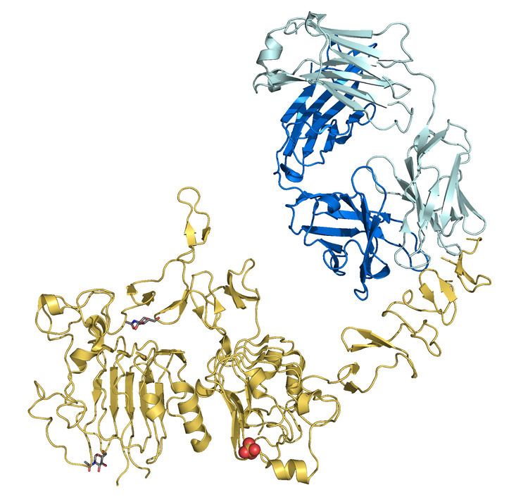
HER2 is the target of the monoclonal antibody trastuzumab (marketed as Herceptin). Trastuzumab is effective only in cancers where HER2 is over-expressed. One year of trastuzumab therapy is recommended for all patients with HER2-positive breast cancer who are also receiving chemotherapy. An important downstream effect of trastuzumab binding to HER2 is an increase in p27, a protein that halts cell proliferation. Another monoclonal antibody, Pertuzumab, which inhibits dimerisation of HER2 and HER3 receptors, was approved by the FDA for use in combination with trastuzumab in June 2012.
As of November 2015, there are a number of ongoing and recently completed clinical trials of novel targeted agents for HER+ metastatic breast cancer, eg. margetuximab.
Additionally, NeuVax (Galena Biopharma) is a peptide-based immunotherapy that directs "killer" T cells to target and destroy cancer cells that express HER2. It has entered phase 3 clinical trials.
It has been found that patients with ER+ (Estrogen receptor positive)/HER2+ compared with ER-/HER2+ breast cancers may actually benefit more from drugs that inhibit the PI3K/AKT molecular pathway.
Over-expression of HER2 can also be suppressed by the amplification of other genes. Research is currently being conducted to discover which genes may have this desired effect.
The expression of HER2 is regulated by signaling through eostrogen receptors. Normally, estradiol and tamoxifen acting through the eostrogen receptor down-regulate the expression of HER2. However, when the ratio of the coactivator AIB-3 exceeds that of the corepressor PAX2, the expression of HER2 is upregulated in the presence of tamoxifen, leading to tamoxifen-resistant breast cancer.
HER2 testing
HER2 testing is performed in breast cancer patients to assess prognosis and to determine suitability for trastuzumab therapy. It is important that trastuzumab is restricted to HER2-positive individuals as it is expensive and has been associated with cardiac toxicity. For HER2-negative tumours, the risks of trastuzumab clearly outweigh the benefits.
HER2 testing on tumour
Tests are usually performed on biopsy samples obtained by either fine-needle aspiration, core needle biopsy, vacuum-assisted breast biopsy, or surgical excision. Immunohistochemistry is used to measure the amount of HER2 protein present in the sample. Alternatively, fluorescence in situ hybridisation (FISH) can be used to measure the number of copies of the gene which are present.
HER2 testing on serum
The extracellular domain of HER2 can be shed from the surface of tumour cells and enter the circulation. Measurement of serum HER2 by enzyme-linked immunosorbent assay (ELISA) offers a far less invasive method of determining HER2 status than a biopsy and consequently has been extensively investigated. Results so far have suggested that changes in serum HER2 concentrations may be useful in predicting response to trastuzumab therapy. However, its ability to determine eligibility for trastuzumab therapy is less clear.
HER2 interactions
HER2/neu has been shown to interact with: