
 | ||
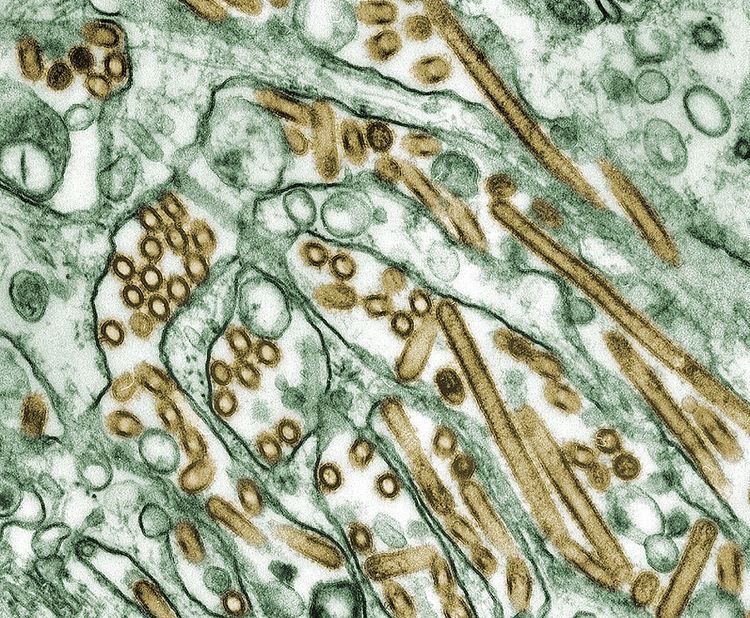
H5N1 genetic structure is the molecular structure of the H5N1 virus's RNA.
Contents
- Terminology
- Context
- Surface encoding gene segments
- Internal encoding gene segments
- Matrix encoding gene segments
- Nucleoprotein encoding gene segments
- Polymerase encoding gene segments
- Mutation
- References
H5N1 is an Influenza A virus subtype. Experts believe it might mutate into a form that transmits easily from person to person. If such a mutation occurs, it might remain an H5N1 subtype or could shift subtypes as did H2N2 when it evolved into the Hong Kong Flu strain of H3N2.
H5N1 has mutated through antigenic drift into dozens of highly pathogenic varieties, but all currently belonging to genotype Z of avian influenza virus H5N1. Genotype Z emerged through reassortment in 2002 from earlier highly pathogenic genotypes of H5N1 that first appeared in China in 1996 in birds and in Hong Kong in 1997 in humans. The "H5N1 viruses from human infections and the closely related avian viruses isolated in 2004 and 2005 belong to a single genotype, often referred to as genotype Z."
This infection of humans coincided with an epizootic (an epidemic in nonhumans) of H5N1 influenza in Hong Kong’s poultry population. This panzootic (a disease affecting animals of many species especially over a wide area) outbreak was stopped by the killing of the entire domestic poultry population within the territory. The name H5N1 refers to the subtypes of surface antigens present on the virus: hemagglutinin type 5 and neuraminidase type 1.
Genotype Z of H5N1 is now the dominant genotype of H5N1. Genotype Z is endemic in birds in southeast Asia and represents a long term pandemic threat.
Influenza A viruses have 11 genes on eight separate RNA molecules [1]:
Two of the most important RNA molecules are HA and PB1. HA creates a surface antigen that is especially important in transmissibility. PB1 creates a viral polymerase molecule that is especially important in virulence.
The HA RNA molecule contains the HA gene, which codes for hemagglutinin, which is an antigenic glycoprotein found on the surface of the influenza viruses and is responsible for binding the virus to the cell that is being infected. Hemagglutinin forms spikes at the surface of flu viruses that function to attach viruses to cells. This attachment is required for efficient transfer of flu virus genes into cells, a process that can be blocked by antibodies that bind to the hemagglutinin proteins.
One genetic factor in distinguishing between human flu viruses and avian flu viruses is that avian influenza HA bind alpha 2-3 sialic acid receptors while human influenza HA bind alpha 2-6 sialic acid receptors. Swine influenza viruses have the ability to bind both types of sialic acid receptors. Humans have avian-type receptors at very low densities and chickens have human-type receptors at very low densities. Some isolates taken from H5N1-infected human have been observed to have HA mutations at positions 182, 192, 223, 226, or 228 and these mutations have been shown to influence the selective binding of the virus to those previously mentioned sialic acid avian and/or human cell surface receptors. These are the types of mutations that can change a bird flu virus into a flu pandemic virus.
A 2008 virulence study that mated in a laboratory an avian flu H5N1 virus that circulated in Thailand in 2004 and a human flu H3N2 virus recovered in Wyoming in 2003 produced 63 viruses representing various potential combinations of human and avian influenza A virus genes. One in five were lethal to mice at low doses. The virus that most closely matched H5N1 for virulence was one with the hemagglutinin (HA), the neuraminidase (NA) and the PB1 avian flu virus RNA molecules with their genes combined with the remaining five RNA molecules (PB2, PA, NP, M, and NS) with their genes from the human flu virus. Both the viruses from the 1957 pandemic and 1968 pandemic carried an avian flu virus PB1 gene. The authors suggest that picking up an avian flu virus PB1 gene may be a critical step in a potential flu pandemic virus arising through reassortment."
PB1 codes for the PB1 protein and the PB1-F2 protein. The PB1 protein is a critical component of the viral polymerase. The PB1-F2 protein is encoded by an alternative open reading frame of the PB1 RNA segment and "interacts with 2 components of the mitochondrial permeability transition pore complex, ANT3 and VDCA1, [sensitizing] cells to apoptosis. [...] PB1-F2 likely contributes to viral pathogenicity and might have an important role in determining the severity of pandemic influenza." This was discovered by Chen et al. and reported in Nature. "After comparing viruses from the Hong Kong 1997 H5N1 outbreak, one amino acid change (N66S) was found in the PB1-F2 sequence at position 66 that correlated with pathogenicity. This same amino acid change (N66S) was also found in the PB1-F2 protein of the 1918 pandemic A/Brevig Mission/18 virus."
Terminology
The Orthomyxovirus family consists of 5 genera: Influenzavirus A, Influenzavirus B, Influenzavirus C, Isavirus, and Thogotovirus.
The "RNA viruses" include the "negative-sense ssRNA viruses" which include the Family "Orthomyxoviridae" which contains five genera, classified by variations in nucleoprotein (NP and M) antigens. One of these is the Genus "Influenzavirus A" which consists of a single species called "Influenza A virus"; one of its subtypes is H5N1.
H5N1 (like the other avian flu viruses) has strains called "highly pathogenic" (HP) and "low-pathogenic" (LP). Avian influenza viruses that cause HPAI are highly virulent, and mortality rates in infected flocks often approach 100%. LPAI viruses are generally of lower virulence, but these viruses can serve as progenitors to HPAI viruses. The current strain of H5N1 responsible for die-offs of domestic birds in Asia is an HPAI strain; other strains of H5N1 occurring elsewhere in the world are less virulent and, therefore, are classified as LPAI strains. All HPAI strains identified to date have involved H5 and H7 subtypes. The distinction concerns pathogenicity in poultry, not humans. Normally a highly pathogenic avian virus is not highly pathogenic to either humans or non-poultry birds. This current strain of H5N1 is unusual in being deadly to so many species.
Both "influenza" (meaning flu) and "A" (meaning species type A) can be used as adjectives of the noun "virus" resulting in the noun phrase "influenza A virus"; which when capitalized is the proper noun Influenza A virus which is the name of the species the noun phrase also refers to.
Context
A virus is one type of microscopic parasite that infects cells in biological organisms.
The Orthomyxoviridae are a family of RNA viruses which infect vertebrates. It includes those viruses which cause influenza. Viruses of this family contain 7 to 8 segments of linear negative-sense single-stranded RNA.
"Influenza virus" refers to a subset of Orthomyxoviridae that create influenza. This taxonomic category is not based on phylogenetics.
Influenza A viruses have 10 genes on eight separate RNA molecules, which, for the reasons mentioned above, are named PB2, PB1, PA, HA, NP, NA, M, and NS. HA, NA, and M specify the structure of proteins that are most medically relevant as targets for antiviral drugs and antibodies. (An eleventh recently discovered gene called PB1-F2 sometimes creates a protein but is absent from some influenza virus isolates.) This segmentation of the influenza genome facilitates genetic recombination by segment reassortment in hosts who are infected with two different influenza viruses at the same time. Influenza A virus is the only species in the Influenzavirus A genus of the Orthomyxoviridae family and are negative sense, single-stranded, segmented RNA viruses.
"The influenza virus RNA polymerase is a multifunctional complex composed of the three viral proteins PB1, PB2 and PA, which, together with the viral nucleoprotein NP, form the minimum complement required for viral mRNA synthesis and replication."
Surface encoding gene segments
Internal encoding gene segments
Matrix encoding gene segments
Nucleoprotein encoding gene segments.
Polymerase encoding gene segments
Mutation
Influenza viruses have a relatively high mutation rate that is characteristic of RNA viruses. The segmentation of the influenza genome facilitates genetic recombination by segment reassortment in hosts who are infected with two different influenza viruses at the same time. H5N1 viruses can reassort genes with other strains that co-infect a host organism, such as a pig, bird, or human, and mutate into a form that can pass easily among humans. This is one of many possible paths to a pandemic.
The ability of various influenza strains to show species-selectivity is largely due to variation in the hemagglutinin genes. Genetic mutations in the hemagglutinin gene that cause single amino acid substitutions can significantly alter the ability of viral hemagglutinin proteins to bind to receptors on the surface of host cells. Such mutations in avian H5N1 viruses can change virus strains from being inefficient at infecting human cells to being as efficient in causing human infections as more common human influenza virus types. This doesn't mean that one amino acid substitution can cause a pandemic, but it does mean that one amino acid substitution can cause an avian flu virus that is not pathogenic in humans to become pathogenic in humans.
H3N2 ("swine flu") is endemic in pigs in China, and has been detected in pigs in Vietnam, increasing fears of the emergence of new variant strains. The dominant strain of annual flu virus in January 2006 was H3N2, which is now resistant to the standard antiviral drugs amantadine and rimantadine. The possibility of H5N1 and H3N2 exchanging genes through reassortment is a major concern. If a reassortment in H5N1 occurs, it might remain an H5N1 subtype, or it could shift subtypes, as H2N2 did when it evolved into the Hong Kong Flu strain of H3N2.
Both the H2N2 and H3N2 pandemic strains contained avian influenza virus RNA segments. "While the pandemic human influenza viruses of 1957 (H2N2) and 1968 (H3N2) clearly arose through reassortment between human and avian viruses, the influenza virus causing the 'Spanish flu' in 1918 appears to be entirely derived from an avian source".
In July 2004, researchers led by H. Deng of the Harbin Veterinary Research Institute, Harbin, China and Professor Robert G. Webster of the St. Jude Children's Research Hospital, Memphis, Tennessee, reported results of experiments in which mice had been exposed to 21 isolates of confirmed H5N1 strains obtained from ducks in China between 1999 and 2002. They found "a clear temporal pattern of progressively increasing pathogenicity". Results reported by Dr. Webster in July 2005 reveal further progression toward pathogenicity in mice and longer virus shedding by ducks.
Asian lineage HPAI A(H5N1) is divided into two antigenic clades. "Clade 1 includes human and bird isolates from Vietnam, Thailand, and Cambodia and bird isolates from Laos and Malaysia. Clade 2 viruses were first identified in bird isolates from China, Indonesia, Japan, and South Korea before spreading westward to the Middle East, Europe, and Africa. The clade 2 viruses have been primarily responsible for human H5N1 infections that have occurred during late 2005 and 2006, according to WHO. Genetic analysis has identified six subclades of clade 2, three of which have a distinct geographic distribution and have been implicated in human infections: Map
A 2007 study focused on the EMA subclade has shed further light on the EMA mutations. "The 36 new isolates reported here greatly expand the amount of whole-genome sequence data available from recent avian influenza (H5N1) isolates. Before our project, GenBank contained only 5 other complete genomes from Europe for the 2004–2006 period, and it contained no whole genomes from the Middle East or northern Africa. Our analysis showed several new findings. First, all European, Middle Eastern, and African samples fall into a clade that is distinct from other contemporary Asian clades, all of which share common ancestry with the original 1997 Hong Kong strain. Phylogenetic trees built on each of the 8 segments show a consistent picture of 3 lineages, as illustrated by the HA tree shown in Figure 1. Two of the clades contain exclusively Vietnamese isolates; the smaller of these, with 5 isolates, we label V1; the larger clade, with 9 isolates, is V2. The remaining 22 isolates all fall into a third, clearly distinct clade, labeled EMA, which comprises samples from Europe, the Middle East, and Africa. Trees for the other 7 segments display a similar topology, with clades V1, V2, and EMA clearly separated in each case. Analyses of all available complete influenza (H5N1) genomes and of 589 HA sequences placed the EMA clade as distinct from the major clades circulating in People's Republic of China, Indonesia, and Southeast Asia."
See http://who.int/csr/disease/avian_influenza/H5CompleteTree.pdf for a Genetic Tree of 1,342 H5N1 viruses based on their HA gene, showing their clade designations.