
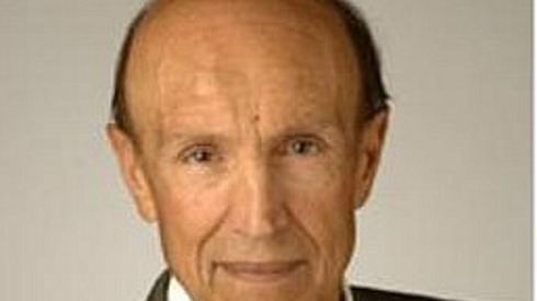
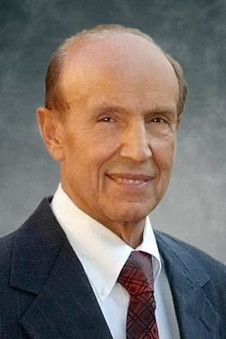
Nationality Iranian-American | Name Gholam Peyman | |
 | ||
Institutions Professor of Basic Medical Sciences at the University of Arizona, Phoenix & Optical Sciences at University of Arizona Tucson, ArizonaEmeritus Professor of Ophthalmology, Tulane University Residence Phoenix, Arizona, United States Books Intravitreal surgery, Vitreous substitutes Fields Ophthalmology, Engineering | ||
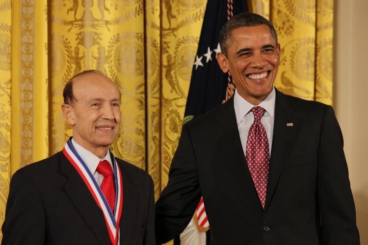
Gholam Peyman - 2011 National Medal of Technology & Innovation
Gholam A. Peyman, MD recipient of National Medal of Technology and Innovation, the nation’s highest honor for technological achievement, bestowed by the President of the United States, President Obama, on America's leading innovators and a Hall of Fame of Ophthalmology and retina surgeon who is also a prolific and successful inventor. Peyman is a member of National Academy of Inventors and has, thus far, been granted 174 US Patents covering a broad range of novel medical devices, intra-ocular drug delivery, surgical techniques, as well as new methods of diagnosis and treatment. His most widely known invention to date is LASIK eye surgery, a vision correction procedure designed to allow people to see clearly without glasses. He was awarded the first US patent for the procedure in 1989 (link to image of patent, below). In addition to the numerous other honors and awards he has received (please see section 5, for Honors and awards), in 2005 he was selected by a ballot among the more than 30,000 ophthalmologists around the world to become one of the thirteen living ophthalmologists inducted into the Ophthalmology Hall of Fame.
Contents
- Gholam Peyman 2011 National Medal of Technology Innovation
- Life and career
- The Invention of LASIK surgery and its improvements
- Other inventions and patents
- Laser in ophthalmology
- Honors and awards
- References
Life and career
Peyman was born in Shiraz, Iran. At the age of 19, he moved to Germany to begin his medical studies. He received his MD at the University of Freiburg in 1962. He completed his internship at St. Johannes Hospital in Diusberg, Germany in 1964 and at Passaic General Hospital in Passaic, New Jersey in 1965. Peyman completed his residency in ophthalmology and a retina fellowship at the University of Essen, Essen Germany, in 1969 and an additional postdoctoral fellowship in retina at the Jules Stein Eye Institute, UCLA School of Medicine in Los Angeles in 1971. Peyman held the position of assistant professor of ophthalmology at the UCLA School of Medicine from 1971 and served as associate professor and then professor of ophthalmology at the Illinois Eye and Ear Infirmary, University of Illinois at Chicago during 1971-1987.
Peyman held a joint appointment at the School of Medicine and also at the Neuroscience Center of Excellence at the Louisiana State University Medical University Medical Center in New Orleans during 1987-2000. During 1998-2000 Peyman held the Prince Abdul Aziz Bin Ahmed Bin Abdul Aziz Al Saud Chair in Retinal Diseases. During 2000-2006, Peyman served as professor of ophthalmology and co-director, Vitreo-Retinal Service, Tulane University School of Medicine in New Orleans. During 2006-2007 he was professor of ophthalmology at the University of Arizona, Tucson, with a cross appointment at University of Arizona College of Optical Sciences. He has been emeritus professor of ophthalmology at Tulane University since 2009. Peyman is currently professor of basic medical sciences at the University of Arizona College of Medicine - Phoenix & Optical engineering at the University of Arizona in Tucson. Peyman was awarded in 2013 an honoree doctorate degree from the National University of Cordoba in Argentina.
The Invention of LASIK surgery and its improvements
At the Illinois Eye and Ear Infirmary, Peyman, because of his interest in the effects of lasers on tissues in the eye, began evaluating the potential use of a CO2 laser to modify corneal refraction in rabbits. No prior study had existed on this concept. The laser was applied to the surface of the cornea using different patterns. This laser created significant scarring. His conclusions at that time were: 1) one has to wait for the development of an ablative laser and 2) one should not ablate the surface of the cornea but, instead, the ablation should take place under a flap in order to prevent scarring, pain and other undesirable sequelae. Peyman published the first article on this subject in 1980.
In late 1982, he read an article from IBM Laboratories, published in Laser Focus, describing the photo-ablative properties of an excimer laser on organic material. This was very exciting information, but, unfortunately, Peyman did not have access to this laser, which at the time was new and very expensive. By 1985 and beyond, many investigators were interested in ablating the corneal surface. However, because of his previous experience with the CO2 laser, Peyman wanted to avoid surface ablation in order to prevent potential corneal scarring and the pain associated with the removal of the corneal epithelium, necessary to expose the surface of the cornea. Therefore, in July 1985, he applied for a patent that described a method of modifying corneal refractive errors using laser ablation under a corneal flap. This US patent was accepted after two revisions and issued in June, 1989. Peyman performed a number of experimental studies evaluating the effect of various excimer lasers in collaboration with Physics Department of the University of Helsinki, Finland. Since he had purchased an Erb-Yag laser in the U.S., he evaluated the concept using this laser in vivo in rabbit and primate eyes and described the creation of a hinged corneal flap to enable the ablation to be performed on the exposed corneal bed, thus reducing the potential for postoperative scarring and pain.
Always aware of the potential limitations of his invention, Peyman devoted considerable time and effort in subsequent years to ameliorating them. In order to improve the risk/benefit considerations of the LASIK procedure, he invented and patented a broad range of ablative and non-ablative inlays to be placed under the surgically created corneal flap. These inlays offered many potential advantages over the standard LASIK technique, the most significant of which is that the inlay procedure is reversible.
However, this procedure has had its own limitations. For example, the implant has been limited to a size of 2 mm diameter and there is also a possibility that a cornea rejects the implant. In 2015 Peyman invented a new refractive surgical procedure that eliminates most of the shortcoming of refractive surgery and implants rejection. His new procedure is now called “Mesik” (Meso, Inside, Implant, Keratomileusis), in which at first a corneal pocket is created with a femtosecond laser, and then the wall of the pocket is crosslinked using riboflavin solution and UV radiation creating a transparent cell-free crosslinked collagen barrier surrounding the implant. This creates an immune privileged cell free space that does not initiate an immune response to an implant. A synthetic organic or polymeric lens can be implanted in the corneal pocket to compensate for the patient’s refractive error. The implant can be exchanged as the eye grows or refractive need dictates.
Other inventions and patents
Peyman's inquiring mind, coupled with his keen interest in engineering and pharmacology along with his in-depth background and experience in ophthalmology resulted, over a 42-year period, in a remarkable range of novel devices, improvements in surgical methods and revolutionary new treatment methods and modalities. A partial list of Peyman's most significant discoveries and inventions (with first publication date), many of which are still in use today, includes:
Eye wall resection (tumors) and biopsy
Glaucoma Pressure-Controlled Shunt
Development of the field of intraocular drug delivery
In the early 1970, Peyman investigated the blood ocular barrier, using peroxidase as a tracer material, to image the site of the barrier in retina which prevented free diffusion of medication from blood in the eye. Because, at that time, despite the systemic antibiotics many eyes were lost after surgery or trauma due to infection, he initiated the evaluation of direct intra-ocular drug delivery. This method has now become an alternative route of drug administration to the systemic and topical medication that in majority of the cases do not cure the infection. Further investigations lead to the prophylactic or therapeutic intraocular administration of the non-toxic doses of numerous medications in the treatment of inflammatory and non-inflammatory diseases of retina in millions of patients. Presently, intraocular drug delivery is accepted and is performed routinely in the treatment of the wet form of age-related macular degeneration and genetic and degenerative diseases of the retina.
Cancer Therapy
Drug delivery
2013:
Laser in ophthalmology
Remote controlled system for Laser Surgery:
Macular degeneration
Artificial Retina Stimulation
Quantum dots and Optogenetic for artificial retinal and brain stimulation and gene therapy
Adaptic optic phoropter for automated vision correction
Honors and awards
1973 Fisher Prize, Chicago Ophthalmology Society
1974 Advisor to the Na.onal Commission of Diabetes
1976 Honorary Member, New Zealand Ophthalmology Society
1979 Honor Award, American Academy of Ophthalmology
1981 Honorary Member, All India Ophthalmological Society
1982 Honorary Member, Paraguayan Ophthalmological Society
1984 Honorary Corresponding Member, Peruvian Ophthalmological Society
1988 Honorary Member, Latin American Ocular Angiofluorography and Photocoagulation Society
1989 U.S. Public Health Service grant EY07541 from the National Eye Institute the National Institutes of Health Services, Bethesda, MD
1989 Senior Honor Award, American Academy of Ophthalmology
1990 Honorary member, All India Ophthalmological Laser Society
1996-97 Included in first edition of The Best Doctors in America: Southeast Region
1997 Honor Award, Vitreous Society
1998 Included in fourth edition of The Best Doctors in America
2001 Gertrude Pyron Lecturer Award, Vitreous Society Annual Mee.ng
2001 ASCRS Innovators Award
2003 Life.me Achievement Award, Iranian Ophthalmology Society
2004 Paul Henkind Lecturer, Macula Society
2005 Hall of Fame of Ophthalmology [Reference: http://www.ascrs.org/Awards/Gholam-A-Peyman-MD.cfm]
2005 Pfizer/ARVO Translational Research Award
2008 Waring Medal, Journal of Refractive Surgery
2008 Lifetime Achievement Award, American Academy of Ophthalmology [REFERENCE: http://www.ascrs.org/Awards/Gholam-A-Peyman-MD.cfm]
2010 Iranian Ophthalmology gold medal
2011 National Highest Medal of Honor in Science and Technology (References: http://www.whitehouse.gov/the-press-office/2012/12/21/president-obama-honors-nation-s-top-scientists-and-innovators; http://www.uspto.gov/about/nmti/recipients/index.jsp)
2013 Distinguished service award UIC department of ophthalmology.
2013 : Health Hero Phoenix Business Journal
2013 : April 14Th Peyman innovation day, Proclaimed by Mayor Stanton Phoenix-AZ
2013: An honoree Doctorate degree from the National University of Cordoba in Argentina.
2013: Elected to the National Academy of Inventors in 2013