
Kingdom Animalia Phylum Platyhelminthes Rank Species Subclass Digenea | Family Paramphistomidae Genus Gastrodiscoides Order Echinostomida | |
 | ||
Similar Watsonius watsoni, Heterophyes heterophyes, Echinostoma ilocanum, Heterophyes, Metagonimus yokogawai | ||
Gastrodiscoides hominis is a zoonotic helminth parasite belonging to a class of flatworms Trematoda. This is the only species in the genus Gastrodiscoides.It is primarily an intestinal fluke of pig, but also infect other vertebrates including human. In fact the first specimen was described from human subject in 1876, and constitute an important parasite of human in Bangladesh, India, Burma, China, Kazakhstan, Philippines, Thailand, Vietnam, and Volga Delta in Russia. In India, it is particularly rampant in Assam, Bengal, Bihar, Madhya Pradesh, Orissa and Uttar Pradesh. Isolated cases are also reported from Africa, such as Nigeria.
Contents
- Discovery
- Description
- Life cycle
- Pathogenicity and pathology
- Epidemiology
- Diagnosis and treatment
- References
Sometimes also known as colonic fluke, it infects the colon of pigs, rhesus monkey, orang-utan, fish, field rats and Napu mouse deer. But in case of humans the worms are attached on the wall of the caecum. Humans are considered an accidental host because the parasite can survive without humans. It causes a helminthic disease called gastrodiscoidiasis.
Discovery
Even though humans are not the natural hosts, G. humanis is unique among helminths because it was first discovered and described from human infection. In 1876, T.R. Lewis and J. McConnell identified from the caecum of an Assamese man in India and named it Amphistomum hominis. Nomenclature was revised by R.T. Leiper in 1913 as Gastrodiscoides humanis, with an invention of the new genus. It was correctly identified in 1939 by J.J.C. Buckley, then a Milner Research Fellow at the London School of Hygiene and Tropical Medicine. His report was the pioneer description of the life cycle of the fluke and prevalence of gastrodiscoidiasis. From a survey of three villages in Assam, he found a surprisingly high incidence, over 40% of the population was infected. It was Buckley more than anyone who was responsible for the formal classification of this parasite.
Description
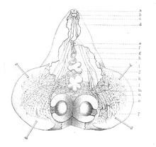
It is a large fluke, somehow vase-shaped and bright-pink in colour. In average it measures 5–8 mm long and 3–5 mm wide. The disc-shaped body is divisible into anterior conical and posterior discoidal regions. The anterior region is a conical projection bearing a prominent oral sucker. The posterior portion is relatively broad (up to 8 mm), discoidal and ventrally excavated. It is an amphistome worm such that the ventral sucker is close to the posterior end. The body covering, tegument is smooth in appearance but the fine structure is actually a series of concentric folds bearing numerous tightly packed tubercles. Ventral surface contains a specialised region of the tegument. Ciliated and non-ciliated papillae are arranged around the oral sucker. The incomplete alimentary canal consists of a pair of lateral pouches arising from the oral sucker and a slightly tortuous pharyngeal tube, which bifurcates into two gut caeca. The large excretory bladder is in the middle behind the ventral sucker. As a hermaphrodite, both male and female reproductive system are present which are arranged in the posterior region. The testes lie in alongside the bifurcation of the caeca, and a common genital pore is on the cone just anterior to the bifurcation. The oval-shaped ovary lies just posterior to the testes in the middle, and the loosely coiled uterus opens to the genital pore. Vitelline glands are scattered around the caeca.
Life cycle
The fluke has a complex life cycle involving at least an intermediate host (presumably aquatic snails), a definitive host (vertebrates), and a free-living stage. Adult worms are found in the lumen of colon (in case of pigs) or caecum (in case of human) from where they discharge hundreds of eggs through self-fertilisation and expel them along the faeces of the host. Egg measures ~146 x 66 μ, and are rhomboidal in shape, transparent, and green in colour. Each egg contains about 24 vitelline cells and a central unembryonated ovum. Eggs in a wet environment hatch into miracidia in 9–14 days. The miracidium grows into the sporocyst stage. It is generally conceived that the unfertilised eggs are ingested by the snail, but there has been no direct observation. In experimental infection of the mollusc Helicorbis coenosus miracidum develops into cercaria after 28–153 days of ingestion. In the snail, mother rediae and daughter rediae are found in the digestive gland, and are about 148–747/45–140 μm in size, sausage-shaped, and lack collar and locomotory organs. The cercariae released from the snail form metacercarial cyst on water plants. However, the complete life cycle is not yet observed in nature and the tiny snail, H. coenosus remains the prime suspect as a vector, as it is coincidentally found in abundance in the pigsties. In some circumstances fishes and other aquatic animals are found to be infected. It is hypothesised that the free cercaria in water bodies, accidentally, find and penetrate these animals as second intermediate host, where they encsyt as metacercaria. These are directly infective to mammals upon consumption or they get attached to vegetation (where night soil is used). Humans ingest the metacercaria either by the infected fish or contaminated vegetable. The parasite travels through the digestive tract into the duodenum then continues down to reach the caecum, where it self-fertilizes and lay eggs, and the cycle continues.
Pathogenicity and pathology
Gastrodiscoidiasis is an infection that is usually asymptomatic and affects the small intestine in animals (such as pigs) to a very mild symptom, but when it occurs in humans it can cause serious health problems and even mortality. It is suspected to cause diarrhoea, fever, abdominal pain, colic, and an increased mucous production. In severe cases, where there are large amounts of eggs present, tissue reactions can occur in the heart or mesenteric lymphatics, and even death may occur if left neglected. Indeed, a number of mortality among Assamese children is attributed to this infection. In pigs, pathological symptoms include infiltration with eosinophils, lymphocytes and plasma cells. The submucosa can show oedema and thickening, resulting in a subacute inflammation of the caecum and mucoid diarrhoea.
Epidemiology
Human gastrodiscoidiasis is endemic in Assam, and to a lesser extent in Philippines. Highest incidence so far recorded is among children in Kamrup district of Assam, where prevalence was as high as 41%. First described from an Indian patient, it was initially believed to have a distribution restricted to India and the southeast Asia. However, investigations reveal that it is widespread, and are further spread by infected persons to other parts of the world, such as Guyana. Level of infection in laboratory animals can be very high among Asian mammals. Regions of high incidence can be attributed to low standard of sanitation, such as rural farms and villages where night soils are used. Infection in both human and animals is most common through the ingestion of vegetation found in contaminated water. It is also assumed that transmission is from infected fish that is under-cooked or eaten in raw, as common among southeast Asian. There is a unique case report of a seven-year-old Nigerian who showed symptoms of malnutrition and anaemia and was eventually diagnosed with infections of G. hominis and Ascaris lumbricoides. The child quickly recovered after proper medication.
Diagnosis and treatment
Diagnosis is made by examination of the faeces and detection of eggs. Adult worms are easily identified from other helminths by their distinctive appearance. Even the eggs alone are readily distinguished from those of other trematodes, by their rhomboid shape and distinct green colour. Patients do not often directly show any symptoms, and if one appears, it indicates that the infection is already at a very high level. There is no prescribed treatment, but traditional practice of soap enema has been very effective in removing the worms. It works to flush the flukes from the colon which removes the parasite entirely since it does not reproduce within the host. Some drugs that have been proven effective are tetrachloroethylene (at a dosage of 0.1 mg/kg on an empty stomach) and the most preferred drug, praziquantel eliminates the parasite with 3 doses (at 25 mg/kg) in one day. Mebendazole was found to be efficient in deworming the parasite from a Nigerian girl who was shedding thousands of parasite eggs in stools even with a single dose of 500 mg. Prevention of this disease is not difficult when simple sanitary measures are taken. Night soil should never be used as a fertilizer because it could contain any number of parasites. Also, vegetables should be washed thoroughly and meat properly cooked.