
 | ||
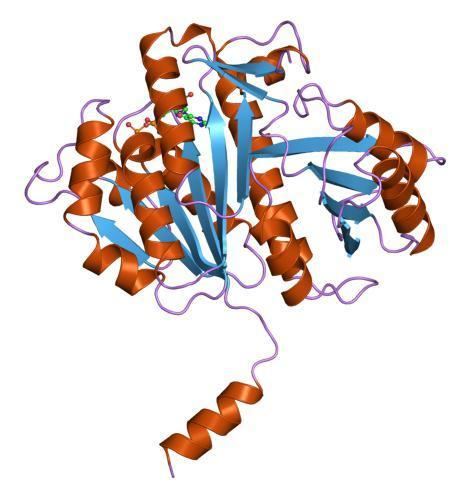
FtsZ is a protein encoded by the ftsZ gene that assembles into a ring at the future site of the septum of bacterial cell division. This is a prokaryotic homologue to the eukaryotic protein tubulin. FtsZ has been named after "Filamenting temperature-sensitive mutant Z". The hypothesis was that cell division mutants of E. coli would grow as filaments due to the inability of the daughter cells to separate from one another.
Contents
History
Discovery of the bacterial cytoskeleton is fairly recent. FtsZ was the first protein of the prokaryotic cytoskeleton to be identified.
The gene was discovered in the 1950s by Y. Hirota (ja:廣田幸敬) and his colleagues in a screen for bacterial cell division mutants. In 1991 it was shown by Erfei Bi and Joseph Lutkenhaus that FtsZ assembled into the Z-ring.
Nuclear-encoded FtsZ in the moss Physcomitrella patens is required for chloroplast division and was the first identified protein essential for organelle division.
Function
During cell division, FtsZ is the first protein to move to the division site, and is essential for recruiting other proteins that produce a new cell wall between the dividing cells. FtsZ's role in cell division is analogous to that of actin in eukaryotic cell division, but, unlike the actin-myosin ring in eukaryotes, FtsZ has no known motor protein associated with it. The origin of the cytokinetic force, thus, remains unclear, but it is believed that the localized synthesis of new cell wall produces at least part of this force. In liposomes Osawa (2009) showed FtsZ is capable of exerting a contractile force with no other proteins present.
Erickson (2009) proposed how the roles of tubulin-like proteins and actin-like proteins in cell division became reversed in an evolutionary mystery. The use of the FtsZ ring in dividing chloroplasts and some mitochondria further establishes their prokaryotic ancestry. It is interesting to note that L-form bacteria that lack a cell wall do not require FtsZ for division, which implies that bacteria may have retained components of an ancestral mode of cell division.
Much is known about the dynamic polymerization activities of tubulin and microtubules, but little is known about these activities in FtsZ. While it is known that single-stranded tubulin protofilaments form into 13 stranded microtubules, the multistranded structure of the FtsZ-containing Z-ring is not known. It is only speculated that the structure consists of overlapping protofilaments.
Recently, proteins similar to tubulin and FtsZ have been discovered in large plasmids found in Bacillus species. They are believed to function as components of segrosomes, which are multiprotein complexes that partition chromosomes/plasmids in bacteria. The plasmid homologs of tubulin/FtsZ seem to have conserved the ability to polymerize into filaments.
The contractile ring
FtsZ has the ability to bind to GTP and also exhibits a GTPase domain that allows it to hydrolyze GTP to GDP and a phosphate group. In vivo, FtsZ forms filaments with a repeating arrangement of subunits, all arranged head-to-tail. These filaments form a ring around the longitudinal midpoint, or septum, of the cell. This ring is called the Z-ring.
The GTP hydrolyzing activity of the protein is not essential to the formation of filaments or division. Mutants lacking the GTPase domain form twisted and disordered septa. These cells with irregular septa can still divide, although abnormally. It is unclear as to whether FtsZ actually provides the physical force that results in division or serves as a marker for other proteins to execute division.
If FtsZ does provide force that divides the cell, it may do so through the relative movement of subunits. Computer models and in vivo measurements suggest that single FtsZ filaments cannot sustain a length more than 30 subunits long. In this model, FtsZ scission force comes from the relative lateral movement of subunits. Lines of FtsZ would line up together parallel and pull on each other creating a "cord" of many strings that tightens itself.
In other models, FtsZ does not provide the contractile force but provides the cell a spatial scaffold for other proteins to execute the division of the cell. This is akin to the creating of a temporary structure by construction workers to access hard-to-reach places of a building. The temporary structure allows unfettered access and ensures that the workers can reach all places. If the temporary structure is not correctly built, the workers will not be able to reach certain places, and the building will be deficient.
The scaffold theory is supported by information that shows that the formation of the ring and localization to the membrane requires the concerted action of a number of accessory proteins. ZipA or the actin homologue FtsA permit initial FtsZ localization to the membrane. Following localization to the membrane, division proteins of the Fts family are recruited for ring assembly. Many of these proteins, such as FtsW, FtsK, and FtsQ are involved in stabilization of the Z ring and may also be active participants in the scission event. The timing of Z-ring formation suggests the possibility of a spatial or temporal signal that permits the formation of FtsZ filaments.
Septal localization and intracellular signaling
The formation of the Z-ring closely coincides with cellular processes associated with replication. Z-ring formation coincides with the termination of genome replication in E. coli and 70% of chromosomal replication in B. subtilis. The timing of Z-ring formation suggests the possibility of a spatial or temporal signal that permits the formation of FtsZ filaments. In Escherichia coli, at least two negative regulators of FtsZ assembly form a bipolar gradient, such that the critical concentration of FtsZ required for FtsZ assembly is lowest at mid-cell between the two segregating chromosomes. This type of regulation seems to occur in other species such as Bacillus subtilis and Caulobacter crescentus. However, other species including Streptococcus pneumoniae and Myxococcus xanthus seem to use positive regulators that stimulate FtsZ assembly at mid-cell.
Communicating distress
FtsZ polymerization is also linked to stressors like DNA damage. DNA damage induces a variety of proteins to be manufactured, one of them called SulA. SulA prevents the polymerization and GTPase activity of FtsZ. SulA accomplishes this task by binding to self-recognizing FtsZ sites. By sequestering FtsZ, the cell can directly link DNA damage to inhibiting cell division.
Preventing DNA damage
Like SulA, there are other mechanisms that prevent cell division that would result in disrupted genetic information sent to daughter cells. So far, two proteins have been identified in E. coli and B. subtilis that prevent division over the nucleoid region: Noc and SlmA. Noc gene knockouts result in cells that divide without respect to the nucleoid region, resulting in its asymmetrical partitioning between the daughter cells. The mechanism is not well understood, but thought to involve sequestration of FtsZ, preventing polymerization over the nucleoid region. SlmA, like SulA, has been observed to sequester FtsZ, preventing the formation of the polymerized Z Ring over the nucleoid region.