
Classification Medical Implant | ||
 | ||
Flexible brain-computer interfaces (fBCIs) are microelectode arrays fabricated on layers of polymers (e.g. polyimide) as seen in flexible electronics 1) to record and process neuronal signaling patterns and 2) to use this data to control an external device. The increased bendability of these devices make them less harmful to brain tissues after immediate implantation and long-term while also helping the arrays access areas of the brain that stiffer silicon-based arrays cannot. Surface, flexible microelectrode arrays can attain resolutions comparable to those of invasive silicon-based arrays and higher than surface silicon-based arrays, making them good candidates for BCIs requiring the ability to resolve signaling patterns on the microscale.
Contents
Brain-Computer Interface Background
Brain-computer interfaces (BCIs) allow communication directly between the brain and external devices, for example letting patients operate an arm remotely or move a wheelchair by thinking about it. This process involves the ability to measure neuronal signaling patterns in vivo in real-time, transmit this data to an external device, analyze the signals using signal processing, and translate the signals obtained into a certain movement.
Origins
The origin of BCIs was the electroencephalogram (EEG). Created in 1924 by the Germany physicist and professor Hans Berger, this device used silver-foil electrodes attached to a patient's scalp to record brain activity via a galvanometer. Today, EEG has become an important diagnostic and research tool in studying patients' neuronal signaling patterns in vivo and can record neuronal signaling with high temporal resolution; it has also been pursued since the 1980s in noninvasive BCI research. Though there have been some successes in using this technology. Though EEGs have potential in BCI applications because of their high temporal resolution, they are limited due to susceptibility to environmental noise and the need to train patients extensively to properly use them.
Microelectrode Arrays and Current BCIs
More widespread is the use of microelectrode arrays in BCIs for recording neuronal signaling. Developed originally in the late 1930s and early 1940s, microelectrodes are usually thin, rigid rods with a sharp tip, allowing individual electrodes to be inserted into the brain and record neuronal signals in real-time from the extracellular or intracellular neuron space; though the electrode shanks were originally made of steel or tungsten, microelectrodes today are usually made from silicon. Penetrating, sub-dural microelectrodes have been organized into arrays since the 1970s to attain signaling patterns from a greater number of neurons and a larger area than can be done by individual micrelectrodes; this includes the well established in vivo Utah and Michigan arrays. However these electrodes can only cover a small surface area of the brain, and microelectrode array technologies for BCIs are currently shifting towards development of surface microelectrode arrays that lay on top of the cortex. Surface microelectrode arrays do not have the high spatial resolution of penetrating arrays; for in vivo use, however, the less harmful effects of surface arrays on the brain is important.
The first major BCI work was done in 1969 by Eberhard Fetz at the University of Washington, in which monkeys controlled deflection of a biofeedback needle with cortical activity. Since, BCI technologies have advanced significantly (especially since the year 2000) with the creation of better algorithms to reconstruct neuronal signaling patterns and advances in microelectronics fabrication techniques. At Duke University, Miguel Nicolelis showed a robotic arm moving in the same pattern as a rhesus monkey reaching for an object on a computer screen due to a BCI reading the monkey's neuronal signaling patterns as it conducted the reaching motion. Taking this a step further, Andrew Schwartz at the University of Pittsburgh showed that rhesus monkeys could feed themselves using a robotic arm controlled by their thoughts; similarly, John Donoghue at Brown University showed that monkeys could move a cursor using their mind to track visual targets on a computer screen. Currently, many of these pioneers in the BCI field have created the private company BrainGate to advance BCI technologies that will allow disabled individuals (e.g. loss of limbs or traumatic spinal cord injury) to conduct everyday functions using thought.
Flexible Electronics for BCIs
Flexible electronics are polymers or other flexible materials (e.g. silk, pentacene, PDMS, parylene, polyimide) that are printed with circuitry; the flexible nature of the organic background materials allowing the electronics created to bend, and the fabrication techniques utilized to create these devices resembles those used to create integrated circuits and microelectromechanical systems (MEMS). Flexible electronics were first developed in 1960s and 1970s, but research interest increased in the mid-2000s. It is expected that flexible electronics will be used in everyday devices ranging from wrist watches to solar cells in the coming decade due to their adaptability. Therefore, it is expected that these devices will be used more widely in the future of fBCI technology.
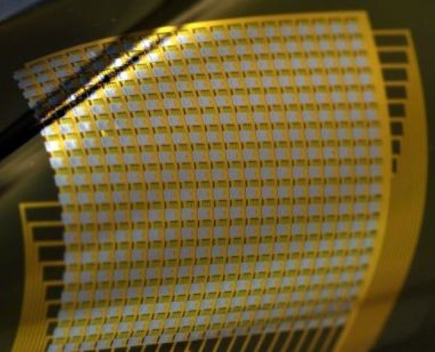
There are currently many fabrication approaches that are being pursued to create efficient, flexible microelectrode arrays. The most notable was for the fBCI created by Jonathan Viventi, now at Duke University, as a graduate student with Brian Litt at the University of Pennsylvania in 2011. This flexible microelectrode array was fabricated using a multi-layer process, with the first layer containing doped silicon ribbons on polyimide, the second layer containing horizontal and vertical interconnects on polyimide, subsequent layers composed of vertical interconnect access structures encapsulated in polyimide or epoxy, and finally platinum contact electrodes. There were 360 individual electrodes of 300 x 300 μm size spaced 500 μm apart in the final array. The polyimide backbone allowed the device to be able to be folded and slid into the sulci or medial areas of the cerebral hemispheres. This array also used multiplexing to efficiently transmit signals recorded in vivo via a wire to an external computer.
Benefits of fBCIs
When interfacing neurons in vitro with microelectrodes, only cytocompatibility is important; this means that the cell contents must not be adversely affected by the electrode. However, when implanting a BCI device into the brain, biocompatibility is necessary; this means that the entire biological system must be compatible with the device. This includes the pressure the device puts on the brain tissues, the cytocompatibility with the neurons, and the long-term scarring effects the device might have on the tissues. Silicon arrays such as the Utah array have been shown in vivo to lack strong biocompatibility. After 6–12 months, these arrays become degraded in the brain and must be replaced; further, long-term use of these arrays can cause hemorrhaging and tissue inflammation. For these reasons, fBCIs show promise of being more biocompatible with the brain in vivo than silicon BCIs.
The lack of biocompatibility of silicon microelectrode arrays with the brain seems to be related to the stiffness of the microelectrodes implanted. Conventional silicon BCIs with Young's moduli of approximately 140 GPa do not deform or conform to brain tissues, which have a Young's modulus of approximately 3.24 kPa (5 orders of magnitude different). This "mechanical mismatch" between brain tissue and the silicon electrodes/microelectrode arrays currently implanted in vivo was further confirmed by Ravi Bellamkonda at Georgia Tech using finite element computational simulations of this system. For this reason, flexible microelectrodes with lower Young's moduli (for example, 3.78 GPa for penetrating flexible microelectrodes created by Michelle LaPlaca at Georgia Tech and 100 kPa for the surface flexible microelectrode array created by Jonathan Viventi) better match the stiffness of the neural environment in which they must exist long-term (ranging from months to decades). The increased bendability of the microelectrode array means not only that the arrays can better conform to the brain and record signals, but that the arrays also do not harm the brain tissue. Lastly, the use of flexible electronics in other biotechnological applications has shown that these systems do not degrade for ten years, meaning that fBCIs are a good long-term alternative to silicon BCIs.
Applications and Current Uses of fBCIs
Flexible electronics have been researched for many biomedical applications, including pressure sensors for stents in blood vessels and electrodes on the heart. Because of this versatility, there have been in recent years many promising applications of flexible microelectrode arrays in the brain. Jonathan Viventi, for example, performed experiments in vivo within feline cat brains to spatially (in a certain space) and temporally (over a certain time) map neuronal signaling patterns (see "Neuronal mapping patterns" section in neurocomputational speech processing). In one experiment, visual evoked responses in the cortex were measured by displaying a white flash of light for 200 ms at various locations on a screen (the visual field); the flexible microelectrode array he created then recorded various patterns of neuronal activity based on the location of the flash on the screen. This data was then used to train a deep belief net (DBN) classifier to identify the location of the flash on the screen, and more recordings were taken to test the trained DBN. Even for the small area (~90 mm^2) of the brain covered, the DBN was able to correctly identify 23 of the 64 screen locations from neuronal recordings, and 42 of 64 locations were identified within 1 neighboring square.
In another experiment, seizures were introduced in the feline brain model using local administration of picrotoxin adjacent to the electrode array on the frontal-medial area of the brain. Because of the device’s ability to mold to this curved portion of the brain and the high spatial resolution of the array, this experiment for the first time mapped spatial patterns of neuronal signaling over time during seizures. It was shown that clockwise and counterclockwise spiraling occurs very rhythmically during picrotoxin-induced epilepsy with the direction of spiraling being changed by various plane waves propagating through the measured area. In addition, it was shown that the spirals detected on the micro-scale were vastly different than the recordings obtained on the macro-scale.
Flexible microelectrode arrays have also been shown to have potential in visual prosthesis technologies, as shown in 2011 by researchers at the Seoul National University. After fabrication, arrow-shaped microelectrodes in an array were placed both inside of agar balls modeling the eyes and in vivo into rabbit eyeballs. The flexible microelectrode array curved and attached to the inside of the eyeballs well, and optical coherence tomography images of the implanted arrays showed good biocompatibility by not tearing the retinal tissue. This first design of a flexible microelectrode array for visual prostheses that fBCIs can be versatile and not only limited to cortical measurements.
Future Aims of fBCI Technology
The implications of fBCIs are vast. One possible clinical implication of this technology is that the fBCI itself may be able to first detect erratic signaling patterns in vivo (such as epileptic micropatterns) that underlie certain neuropathologies and then counteract these patterns by sending their own "cancelling" electrical signals.
The high resolution provided by this sub-dural flexible microelectrode array would also be able to adequately measure brain activity patterns spatially and temporally for applications in BCI systems without loss of device performance or injuring brain tissue in the long-term. Expanding the device to cover larger areas of the brain or using more arrays to cover more parts of the brain would allow for more holistic analysis of neural signaling, thus creating more patient control and confidence in performing tasks using an external device. In addition, it may also be possible for patients to control multiple devices that serve various purposes simultaneously using fBCIs implanted in various regions of the brain.
Lastly, flexible recording arrays would be able to be implanted between the two hemispheres of the brain, allowing BCI devices, for example, to record information from the motor cortex on both sides of the brain; this signaling could then be used to implement movements requiring multiple artificial prosthetic limbs. Further, the flexible nature of fBCIs implies that other implanted electrode technologies, such as the deep brain stimulation (DBS) electrode that is implanted to suppress tremors in Parkinson's and essential tremor patients, may be able to conform and bend to important areas of the brain for recording more efficiently. For these reasons, the creation of fBCI technologies is an emerging field with major implications for how the human body will interface with external devices in the near future.