
Family †Akidnognathidae Rank Genus | Phylum Chordata Order Therapsid | |
 | ||
Similar Therocephalia, Megawhaitsia, Therapsid, Moschorhinus, Ericiolacerta | ||
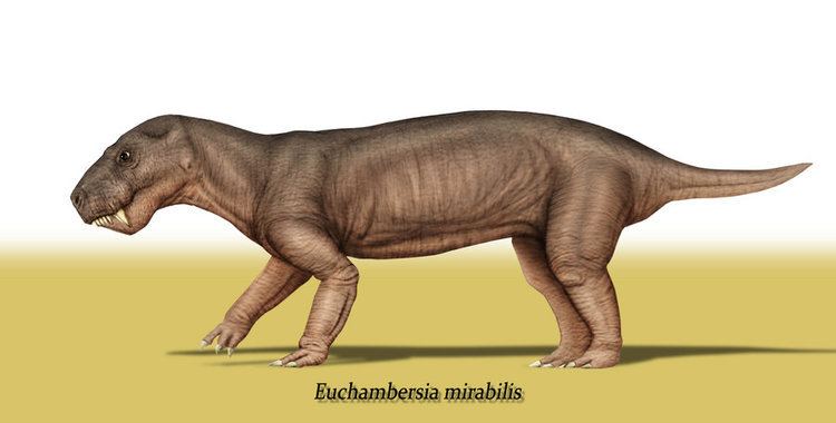
Euchambersia is a genus of therocephalian therapsid that lived during the Late Permian, approximately 255 million years ago, in what is now South Africa. The genus contains a single species, E. mirabilis, named by paleontologist Robert Broom in 1931 from a skull missing the lower jaws; a second skull, belonging to an immature individual, was later described. It is a member of the family Akidnognathidae, which historically has also been referred by as the synonymous Euchambersiidae (named after Euchambersia).
Contents
- Description
- Teeth
- Maxillary fossa and associated canals
- Discovery and naming
- Classification
- Venom
- Paleoecology
- In popular culture
- References
Euchambersia is notable among therocephalians for possessing ridged canines and a large indentation in the side of the skull. Under the erroneous assumption that the canines are grooved instead of ridged, it has been proposed that these structures supported a venom delivery mechanism. More recently, the internal structure of the skull of Euchambersia has been used as stronger evidence in favour of the hypothesis that it was venomous; other possibilities, such as the indentation supporting some sort of sensory organ, still remain plausible.
Description
Euchambersia was small and short-snouted for a therocephalian, with the type skull having a length of approximately 116 millimetres (4.6 in) once crushing and deformation of the fossil was reconstructed. The second skull belongs to a smaller individual, with a length of 80 millimetres (3.1 in); it was probably immature, judging by the lack of fusion in the skull.
Teeth
Although the skulls are incompletely preserved, CT scanning suggests that each premaxilla held five incisors, with the sockets becoming progressively larger from the first to the fifth incisor. Like other theriodonts, the crowns of the incisors are conical; they also lack serrations, unlike gorgonopsians and scylacosaurian therocephalians. The interior edge of the incisors seems to be slightly concave, and the back edge appears to have a ridge. The smaller specimen has an incisor preserved within its nasal cavity; it is more strongly recurved and has wear marks on its top edge, suggesting that it is probably a lower incisor. Its fourth incisor also has a replacement tooth growing behind it, accompanied by resorption of the root.
The type specimen prominently preserves the right canine. It is large and round in cross-section, and bears a prominent ridge on the side of its front surface. Immediately beside this ridge is a shallow depression that becomes wider near the top of the tooth, which may have been interpreted as a groove by some authors. Theriodonts usually replace their teeth in an alternating (or distichial) pattern, such that the canine tooth is always functional; both skulls of Euchambersia show no sign of any replacement teeth developing, suggesting that Euchambersia was reliant on having both canines present and functional simultaneously.
Maxillary fossa and associated canals
Behind the incisors and canines, there were no additional teeth in the jaw. Where teeth would be located in therocephalians that do have teeth behind the canines, there is instead a large depression, or fossa, in side of the maxilla. This fossa is 48% the length of the jaw in the type specimen, and 38% in the second skull. In both skulls, this fossa is divided into two parts: a shallower ridge on top, and a larger and deeper depression on the bottom. A wide furrow beginning behind the canine contacts the bottom of the fossa and then passes into the interior of the mouth. The bottom portion of the fossa is strongly pitted and bears a small opening, or foramen, on both the front and back surfaces.
CT scanning shows that these openings lead to canals that connect to the trigeminal nerve, which controls facial sensitivity. The forward-directed canal also splits into the three main branches of the infraorbital nerve, all of which connect to the socket of the canine; the junction occurs about 3–6 millimetres (0.12–0.24 in) along the canal, another point of variation between the two skulls. The top branch, the external nasal ramus, splits into four branches in the type skull, but it does not split in the second skull. In other therapsids like Thrinaxodon, Bauria, and Olivierosuchus, the external nasal ramus generally splits into three or more branches. All of these canals would have brought nerves and nutrient-rich tissue to the root of the canines and the rest of the upper jaw.
Discovery and naming
The type specimen of Euchambersia was found on the South African farm of Vanwyksfontein, near the town of Colesberg. It consists of a single, distorted skull, catalogued as NHMUK R5696 and described by Robert Broom in 1931. A second, smaller skull, with the specimen number BP/1/4009, was later found in 1966 and described by Kitching in 1977. Both came from the same general layer of rock, in the Cistecephalus assemblage zone of the Beaufort Group within the Karoo Supergroup. This layer of rock has been dated to the Wuchapingian stage of the Late Permian, between 256.2 and 255.2 Mya.
Classification
In 1934, Euchambersia was assigned to the family Euchambersiidae by Boonstra. This family is a junior synonym of the Akidnognathidae, since Akidnognathus (which also belongs in the same family) was named prior to Euchambersia. In 2008, Ivakhnenko included the Akidnognathidae (under the name Euchambersiidae) as the sister group of the family Whaitsiidae in the superfamily Whaitsioidea. However, more recent phylogenies do not include the Akidnognathidae in the Whaitsioidea; the phylogeny of Huttenlocker and Sidor (2016) is shown below.
Venom
The large maxillary fossae of Euchambersia has been a continual subject of debate regarding its function, but most researchers agree that it held some sort of secretory gland. While Broom initially argued that the fossae may have contained the parotid salivary glands, this proposal was rejected by Boonstra and Lehman, who noted that the parotid glands tend to be placed behind the eye; they respectively suggested that the fossae held modified lacrimal glands and Harderian glands. However, the latter is also unlikely because Harderian glands are usually placed inside the eye socket. Nopcsa suggested that the maxillary fossae housed venom glands (which may have been derived from lacrimal glands), with the ridged canines and the notches behind the canines allowing the venom to flow passively into the victim's bloodstream. This hypothesis was widely accepted throughout the 20th century and the characteristic morphology of Euchambersia was used to support possible venom-bearing adaptations among various other prehistoric animals, including the therocephalian Ichibengops.
Much of this acceptance has been based on the erroneous assumption that the canines are grooved instead of ridged; grooved canines in Euchambersia would parallel the fangs of various venomous snakes as well as the venom-delivering incisors of solenodons. This interpretation, which has consistently appeared in literature published after 1986, was subsequently determined to be the result of the propagation of Broom's overly reconstructed diagram of the skull, without the context of the actual specimens. This line of evidence has been raised to support the necessity of a re-evaluation the hypothesis of a venomous bite in Euchambersia. Additionally, it has also been argued that grooved and ridged canines are not necessarily associated with venomous animals either, as shown by their presence in hippopotami, muntjacs, and baboons, in which they play a role in grooming or sharpening the teeth; in the latter two, ridged canines are also accompanied by a distinct fossa in front of the eye, which is entirely unconnected with venom. Furthermore, grooved and ridged teeth in non-venomous snakes are used to reduce suctional drag when capturing slippery prey like fish or invertebrates.
CT scanning of the known specimens of Euchambersia were subsequently used to provide more concrete support in favour of the venom hypothesis. The canals leading into and from the maxillary fossae, which were revealed by the scans, would primarily have supported the trigeminal nerve as well as blood vessels. However, the fact that the canals also directly lead to the root of the canines would suggest that they had a secondary role in venom delivery. In all, Euchambersia seems to have had a venom gland (housed in the maxillary fossae), a delivery mechanism of the venom (the maxillary canals), and an instrument by which a wound for venom delivery can be inflicted (the ridged canines), which satisfy the criteria of a venomous animal. However, this does not conclusively demonstrate that Euchambersia was actually venomous, especially given the previously stated objections. Additionally, there are no living animals with a delivery system analogous to the proposed system for Euchambersia (most deliver venom through the lower jaw, while snakes have specialized ducts).
An alternate hypothesis suggested by the CT scans involves some kind of sensory organ occupying the maxillary fossa. Uniquely among therapsids, the canal within the maxilla is exposed on the back side of the maxillary fossa, which implies that the canal, carrying the trigeminal nerve, would probably have extended across the fossa, outside of the outline of the skull. This may have supported a specialized sensory organ analogous to the pit organ of pit vipers and some other snakes, or alternatively a ganglion of nerve cells. It is also possible that this organ functioned as a replacement for the parietal eye in Euchambersia, like the pit organ does in pit vipers. However, such an expanded sensory organ would be unprecedented among tetrapods, and the few other therocephalians that also lack a parietal eye do not have a maxillary fossa either. Thus, the venom hypothesis is more plausible.
Paleoecology
The Cistecephalus Assemblage Zone, from where Euchambersia is known, represents a floodplain that was covered in many small, relatively straight streams. The water level in these streams appeared to have been seasonally dependent.
In the Cistecephalus AZ, other co-occurring therocephalians included Hofmeyria, Homodontosaurus, Ictidostoma, Ictidosuchoides, Ictidosuchops, Macroscelesaurus, Polycynodon, and Proalopecopsis. More numerous, however, were the gorgonopsians, which included Aelurognathus, Aelurosaurus, Aloposaurus, Arctognathus, Arctops, Cerdorhinus, Clelandina, Cyonosaurus, Dinogorgon, Gorgonops, Lycaenops, Leontocephalus, Pardocephalus, Prorubidgea, Rubidgea, Scylacops, Scymnognathus, and Sycosaurus.
By far the most abundant herbivore was the dicynodont Diictodon, with over 1900 known specimens from the Cistecephalus AZ. Other dicynodonts included Aulacephalodon, Cistecephalus, Dicynodon, Dicynodontoides, Digalodon, Dinanomodon, Emydops, Endothiodon, Kingoria, Kitchinganomodon, Oudenodon, Palemydops, Pelanomodon, Pristerodon, and Rhachiocephalus. The biarmosuchians Lemurosaurus, Lycaenodon, Paraburnetia, and Rubidgina were also present, along with the cynodonts Cynosaurus and Procynosuchus. Non-synapsids included the archosauromorph Younginia; the parareptilians Anthodon, Milleretta, Nanoparia, Owenetta, and Pareiasaurus; and the temnospondyl Rhinesuchus.
In popular culture
The venomous bite of Euchambersia, widely accepted at the time, was the basis of a sequence involving venomous therocephalians in the BBC documentary Walking with Monsters.