
 | ||
Electron density is the measure of the probability of an electron being present at a specific location.
Contents
In molecules, regions of electron density are usually found around the atom, and its bonds. In de-localized or conjugated systems, such as phenol, benzene and compounds such as hemoglobin and chlorophyll, the electron density covers an entire region, i.e., in benzene they are found above and below the planar ring. This is sometimes shown diagrammatically as a series of alternating single and double bonds. In the case of phenol and benzene, a circle inside a hexagon shows the de-localized nature of the compound. This is shown below:
In compounds with multiple ring systems which are interconnected, this is no longer accurate, so alternating single and double bonds are used. In compounds such as chlorophyll and phenol, some diagrams show a dotted or dashed line to represent the de-localization of areas where the electron density is higher next to the single bonds. Conjugated systems can sometimes represent regions where electromagnetic radiation is absorbed at different wavelengths resulting in compounds appearing coloured. In polymers, these areas are known as chromophores.
In quantum chemical calculations, the electron density, ρ(r), is a function of the coordinates r, defined so ρ(r)dr is the number of electrons in a small volume dr. For closed-shell molecules,
where P is the density matrix. Electron densities are often rendered in terms of an isosurface (an isodensity surface) with the size and shape of the surface determined by the value of the density chosen, or in terms of a percentage of total electrons enclosed.
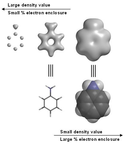
Molecular modeling software often provides graphical images of electron density. For example, in aniline (see image at right). Graphical models, including electron density are a commonly employed tool in chemistry education. Note in the left-most image of aniline, high electron densities are associated with the carbons and nitrogen, but the hydrogens with only one proton in their nuclei, are not visible. This is the reason that X-ray diffraction has a difficult time locating hydrogen positions.
Most molecular modeling software packages allow the user to choose a value for the electron density, often called the IsoValue. Some software also allows for specification of the electron density in terms of percentage of total electrons enclosed. Depending on the IsoValue (typical units are electrons per cubic bohr), or the percentage of total electrons enclosed, the electron density surface can be used to locate atoms, emphasize electron densities associated with chemical bonds, or to indicate overall molecular size and shape.
Graphically, the electron density surface also serves as a canvas upon which other electronic properties can be displayed. The electrostatic potential map (the property of electrostatic potential mapped upon the electron density) provides an indicator for charge distribution in a molecule. The local ionization potential map (the property of local ionization potential mapped upon the electron density) provides an indicator of electrophilicity. And the LUMO map (lowest unoccupied molecular orbital mapped upon the electron density) can provide an indicatory for nucleophilicity.
Experiments
Many experimental techniques can measure electron density. For example, through X-ray diffraction scans, where X-rays of a suitable wavelength are targeted towards a sample and measurements are made over time, gives a probabilistic representation of where electrons can be found. From these positions molecular structures can often be determined for crystallized systems. Quantum electrodynamics and some branches of quantum theory also study and analyze electron superposition and other related phenomena. Mulliken population analysis is based on electron densities in molecules and is a way of dividing the density between atoms to give an estimate of atomic charges.
In transmission electron microscopy (TEM) and deep inelastic scattering, as well as other high energy particle experiments, high energy electrons inderacts with the electron cloud to give a direct representation of the electron density. TEM, scanning tunneling microscopy (STM) and atomic force microscopy (AFM) can be used to probe the electron density of specific individual atoms.
Spin density
Spin density is electron density applied to free radicals. It is defined as the total electron density of electrons of one spin minus the total electron density of the electrons of the other spin. One of the ways to measure it experimentally is by electron spin resonance, neutron diffraction allows direct mapping of the spin density in 3D-space.