
 | ||
Elastofibroma, also called elastofibroma dorsi, is an ill-defined fibroelastic tumor-like condition made up of enlarged and irregular elastic fibers.
Contents
Origin
There are several theories about origin:
Signs and Symptoms
Patient will present with a slow growing, deep-seated, firm mass, often presenting bilaterally. There may be pain or tenderness, but this is rare.
Imaging findings
By computed tomography, there is a poorly circumscribed, heterogeneous soft tissue mass, with a signal intensity similar to skeletal muscle. The fact that the lesion may be bilateral, helps eliminate a sarcoma from further consideration. At US, elastofibromas are depicted deep to the musculature as a multilayered pattern of hypoechoic linear areas of fat deposition intermixed with echogenic fibroelastic tissue. The mass often protrudes from the subscapular region upon shoulder abduction, allowing better delineation of the finding.
Pathology findings
In general, the tumor is an ill defined, nonencapsulated, rubbery, and firm, white lesion with interspersed fat. The tumors can be quite large (up to 20 cm), although most are around 5 cm.
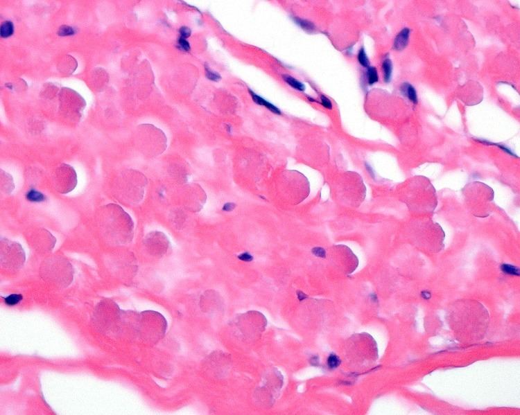
By microscopie view, there is an admixture of heavy dense bands of collagenous tissue dissected by fat and abnormal elastic fibers. The elastic fibers are often quite large and are easily identified. The elastic fibers are coarse, thick, and darkly eosinophilic, often fragmented into globules, creating a "string of pearls" or "pipe cleaner" appearance. Because of degeneration, the elastic fibers will appear as globules with a serrated or "prickled" edge.
Histochemistry
The elastic fibers will be highlighted by a Weigert or von Gieson elastic stains.
Differential diagnoses
Given the anatomic site, a spindle cell lipoma, nuchal-type fibroma and fibromatosis colli are all included in the differential diagnosis.
Management
Simple excision is the treatment of choice, although given the large size, bleeding into the space can be a potential complication. Isolated recurrences may be seen, but there is no malignant potential.
Epidemiology
This is a very rare phenomenon (< 0.001% of soft tissue tumors), usually presenting in elderly patients (>50 years of age), and more commonly in women than men (5:1). There is an increased frequency in Okinawa, Japan, but this may be a reporting bias. The tumor develops very specifically in the subscapular or infrascapular area, deep to the muscle, sometimes even attached to periosteum of ribs. It is usually between the shoulder blade and the lower neck, with rare tumors reported in the chest wall.