
 | ||
Diamond nanoparticles, also known as nanodiamonds, are single crystal diamonds that range anywhere from 5 to 500 nm. Because of their inexpensive, large-scale synthesis, potential for surface functionalization, and high biocompatibility, nanodiamonds are widely investigated as a potential material in biological and electronic applications and quantum engineering.
Contents
Structure and composition
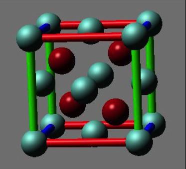
There are three main aspects in the structure of diamond nanoparticles to be considered: the overall shape, the core, and the surface. Through multiple diffraction experiments, it has been determined that the overall shape of diamond nanoparticles is either spherical or elliptical. At the core of diamond nanoparticles lies a diamond cage, which is composed mainly of carbons. While the core closely resemble the structure of a diamond, the surface of diamond nanoparticles actually resemble the structure of graphite. A recent study shows that the surface consists mainly of carbons, with high amounts of phenols, pyrones, and sulfonic acid, as well as carboxylic acid groups, hydroxyl groups, and epoxide groups, though in lesser amounts. Occasionally, defects such as nitrogen-vacancy centers can be found in the structure of diamond nanoparticles. 15N NMR research confirms presence of such defects. A recent study shows that the frequency of nitrogen-vacancy centers decreases with the size of diamond nanoparticles.
Production methods
Nanodiamond synthesis was first discovered in the USSR in 1963 when Russian researchers found that the detonation of carbon-based explosives resulted in the formation of nanodiamonds. Other methods of synthesis include hydrothermal synthesis, ion bombardment, laser bombardment, microwave plasma chemical vapor deposition techniques, ultrasound synthesis, and electrochemical synthesis. In addition, the decomposition of graphitic C3N4 under high pressure and high temperature yields large quantities of high purity diamond nanoparticles. However, detonation synthesis of nanodiamonds has become the industry standard in the commercial production of nanodiamonds: the most commonly utilized explosives being mixtures of trinitrotoluene and hexogen or octogen. Detonation is often performed in a sealed, oxygen-free, stainless steel chamber and yields a mixture of nanodiamonds averaging 5 nm and other graphitic compounds. In detonation synthesis, nanodiamonds form under pressures greater than 15 GPa and temperatures greater than 3000K in the absence of oxygen to prevent the oxidation of diamond nanoparticles. The rapid cooling of the system increases nanodiamond yields as diamond remains the most stable phase under such conditions. Detonation synthesis utilizes gas-based and liquid-based coolants such as argon and water, water-based foams, and ice. Because detonation synthesis results in a mix of nanodiamond particles and other graphitic carbon forms, extensive cleaning methods must be employed to rid the mixture of impurities. In general, gaseous ozone treatment or solution-phase nitric acid oxidation is utilized to remove sp2 carbons and metal impurities.
Applications
Diamond nanoparticles have the potential to be utilized in a myriad of biological applications and due to their unique properties such as inertness and hardness, nanodiamonds may prove to be a better alternative to the traditional nanomaterials currently utilized to carry drugs, coat implantable materials, and synthesize biosensors and biomedical robots. The low cytotoxicity of diamond nanoparticles affirms their utilization as biologically compatible materials.[ In vitro studies exploring the dispersion of diamond nanoparticles in cells have revealed that most diamond nanoparticles exhibit fluorescence and are uniformly distributed. Further studies have been conducted to decrease the size of nanodiamonds and functionalize their surface. Surface-modified diamond nanoparticles can be used as highly effective antigen delivery carriers, which suggests that modified nanodiamonds have immense potential to be utilized as an alternative to traditional catalysts. Studies have shown that small photoluminescent diamond nanoparticles that remain free in the cytosol are excellent contenders for the transport of biomolecules. In addition, larger sized nanodiamonds, due to their “high uptake efficiency,” have the potential to serve as cellular labels. Studies have concluded that diamond nanoparticles are similar to carbon nanotubes and upon being treated with surfactants, the stability and biocompatibility of both carbon nanotubes and the nanodiamonds in solution greatly increase. In addition, the ability to surface functionalize nanodiamonds of small diameters provides various possibilities for diamond nanoparticles to be utilized as biolabels with potentially low cytotoxicity.