
Symbol Apocytochr_F_C Pfam clan CL0105 PROSITE PDOC00169 | Pfam PF01333 InterPro IPR002325 SCOP 1ctm | |
 | ||
Cytochrome f is the largest subunit of cytochrome b6f complex (plastoquinol—plastocyanin reductase; EC 1.10.99.1). In its structure and functions, the cytochrome b6f complex bears extensive analogy to the cytochrome bc1 complex of mitochondria and photosynthetic purple bacteria. Cytochrome f (cyt f) plays a role analogous to that of cytochrome c1, in spite of their different structures.
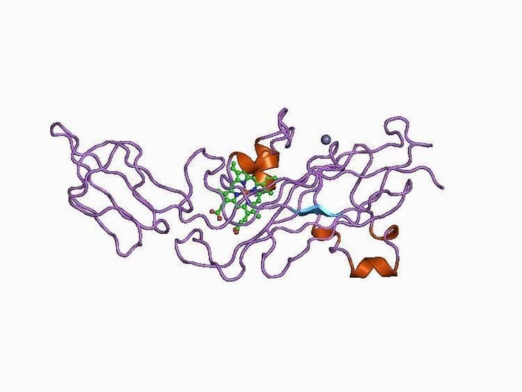
The 3D structure of Brassica rapa (Turnip) cyt f has been determined. The lumen-side segment of cyt f includes two structural domains: a small one above a larger one that, in turn, is on top of the attachment to the membrane domain. The large domain consists of an anti-parallel beta-sandwich and a short haem-binding peptide, which form a three-layer structure. The small domain is inserted between beta-strands F and G of the large domain and is an all-beta domain. The haem nestles between two short helices at the N terminus of cyt f. Within the second helix is the sequence motif for the c-type cytochromes, CxxCH (residues 21-25), which is covalently attached to the haem through thioether bonds to Cys-21 and Cys-24. His-25 is the fifth haem iron ligand. The sixth haem iron ligand is the alpha-amino group of Tyr-1 in the first helix. Cyt f has an internal network of water molecules that may function as a proton wire. The water chain appears to be a conserved feature of cyt f.