
 | ||
A cortical homunculus is a physical representation of the human body, located within the brain. A cortical homunculus is a neurological "map" of the anatomical divisions of the body. There are two types of cortical homunculi: sensory and motor.
Contents
Types
The primary motor cortex is located in the precentral gyrus, and handles signals coming from the premotor area of the frontal lobes. The primary sensory cortex is located in the postcentral gyrus, and handles signals coming from the thalamus. These signals are then transmitted from the gyri to the brain stem and spinal cord via corresponding nerves.
Motor
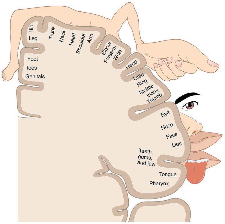
At the primary motor cortex, motor representation is arranged in an orderly manner, inverted. The toes are represented at the top of the cerebral hemisphere, while the mouth is represented at the bottom of the hemisphere, closer to the lateral sulcus. These representations lie along a fold in the cortex called the central sulcus. The homunculus is split in half, with motor representation for each side of the body represented on the opposite side of the brain. Some body parts may be controlled by partially overlapping regions of cortex.
The amount of cortex devoted to any given body region is proportional to how richly innervated that region is, not to the body region's physical size. Areas of the body with greater or more complex sensory or motor connections are represented as larger in the homunculus, those with fewer or less complex connections are represented as smaller. The resulting image is that of a distorted human body, with disproportionately huge hands, lips, and face.
Development
Dr. Wilder Penfield used a similar image to depict the body according to the areas of the motor cortex controlling it in voluntary movement. Sometimes thought to be the brain's map of the body, the motor homunculus really is a map of the proportionate association of the cortex with body members. It also reflects kinesthetic proprioception, the body as felt in motion.
Penfield's motor homunculus is usually shown as a canonical, 2-D diagram. This is an oversimplification, as it does not show the original data Penfield collected from patients undergoing surgery for epilepsy. This simplification suggests that lesions of the motor cortex will give rise to specific deficits in specific muscles. This misconception is not necessarily the case: lesions produce deficits in groups of synergistic muscles. This difference is critical, as it suggests that the motor cortex functions in terms of overall movements as entire libraries, rather than how to move individual muscles, allowing a great deal more complexity.
Dr. Wilder Penfield Sensory & Motor Homunculus
Penfield's 3 dimensional homunculus is an illustration of the sensory and motor representation of the body within the brain or the cortex man. These figures represent perhaps the most famous conceptual map in modern neuroscience.
Penfield first conceived his Homunculus as a thought experiment in order to conceptualize and objectify human brain function. Penfield's experiment went so far as to create an imaginary world, in which his Homunculus lived, the world of 'if'.
Penfield referred to his creations as ‘grotesque creatures' which illustrate the highly suggestive concept of the body within the brain or the cortex man. Crucial to Penfield's Homunculi, was the fact that it's body parts developed in proportion to the surface area of the cortical region dedicated to the control of specific functions.
Penfield's Homunculus looked like strange grotesque creatures. For example, the afferent sensory nerves arriving from the hands, terminate over a large areas of the brain, this results in the hands of the homunculus being correspondingly large. In contrast, the nerves emanating from the torso or arms, cover a much smaller area, consequently, the torso and arms of the homunculus look small and weak.
Importantly, Penfield was not the first person to attempt to objectify human brain function through the means of a Homunculus. What made Penfields different however, was that he recognized the intersection of sensory and motor function. Penfield treated the two parts of the reflex arc as being distinct and separate. He experimented and tested separately, for both sensory and motor function. This experimental work created a division between the sensory and motor investigations, a division that resulted in the structuring principles of Penfield's work. Work which would ultimately radically change the parameters of neurological investigation.
Penfield's Homunculus come in two forms, the Sensory and Motor Homunculus. Perhaps the most well known of Penfield's Homunculus, are the pair on permanent display at the Natural History Museum, London.