
Dorlands/Elsevier f_02/12354419 FMA 20275 | TA A04.5.01.020 | |
 | ||
Latin falx inguinalis, tendo conjunctivus | ||
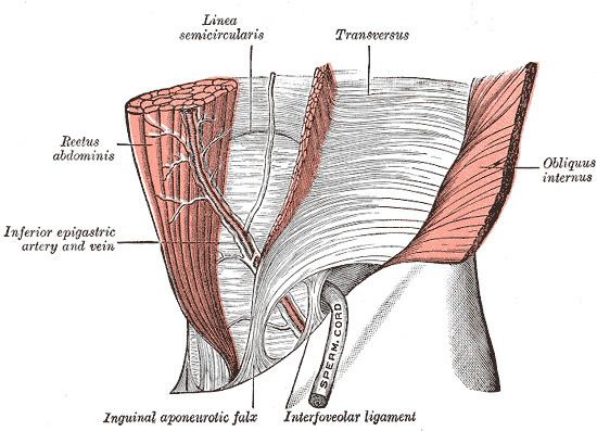
The conjoint tendon (previously known as the inguinal aponeurotic falx) is a structure formed from the lower part of the common aponeurosis of the internal abdominal oblique and the transverse abdominal as it inserts into the crest of the pubis and pectineal line immediately behind the superficial inguinal ring. It is usually conjoint with the tendon of the abdominal internal oblique muscle, but they may be separate as well. It forms the medial part of the posterior wall of the inguinal canal.
Clinical significance
The conjoint tendon serves to protect what would otherwise be a weak point in the abdominal wall. A weakening of the conjoint tendon can precipitate a direct inguinal hernia.
A direct inguinal hernia will protrude through Hesselbach's triangle, whose borders are the rectus abdominus (medially), inferior epigastric artery and vein (superolaterally), and the inguinal ligament (inferiorly). The hernia will lie medial to the inferior epigastric artery. This is in contrast to an indirect inguinal hernia, which will protrude laterally to the inferior epigastric artery and is most commonly due to an embryological defect in the closure of the deep inguinal ring.