
Dorlands
/Elsevier n_05/12565792 | TA A14.2.07.047 | |
 | ||
From sacral plexus via sciatic nerve (L4-S2) To Deep fibular nerve and Superficial fibular nerve Innervates Anterior compartment of leg, lateral compartment of leg, extensor digitorum brevis Latin Nervus fibularis communis,
Nervus peronaeus communis | ||
The common peroneal nerve (common fibular nerve; external popliteal nerve; lateral popliteal nerve) is a nerve in the lower leg that provides sensation and motor function to parts of the lower leg. When damaged or compressed, it can cause foot drop.
Contents
Structure
The common peroneal nerve, about one-half the size of the tibial nerve, arises from the dorsal branches of the fourth and fifth lumbar and the first and second sacral nerves.
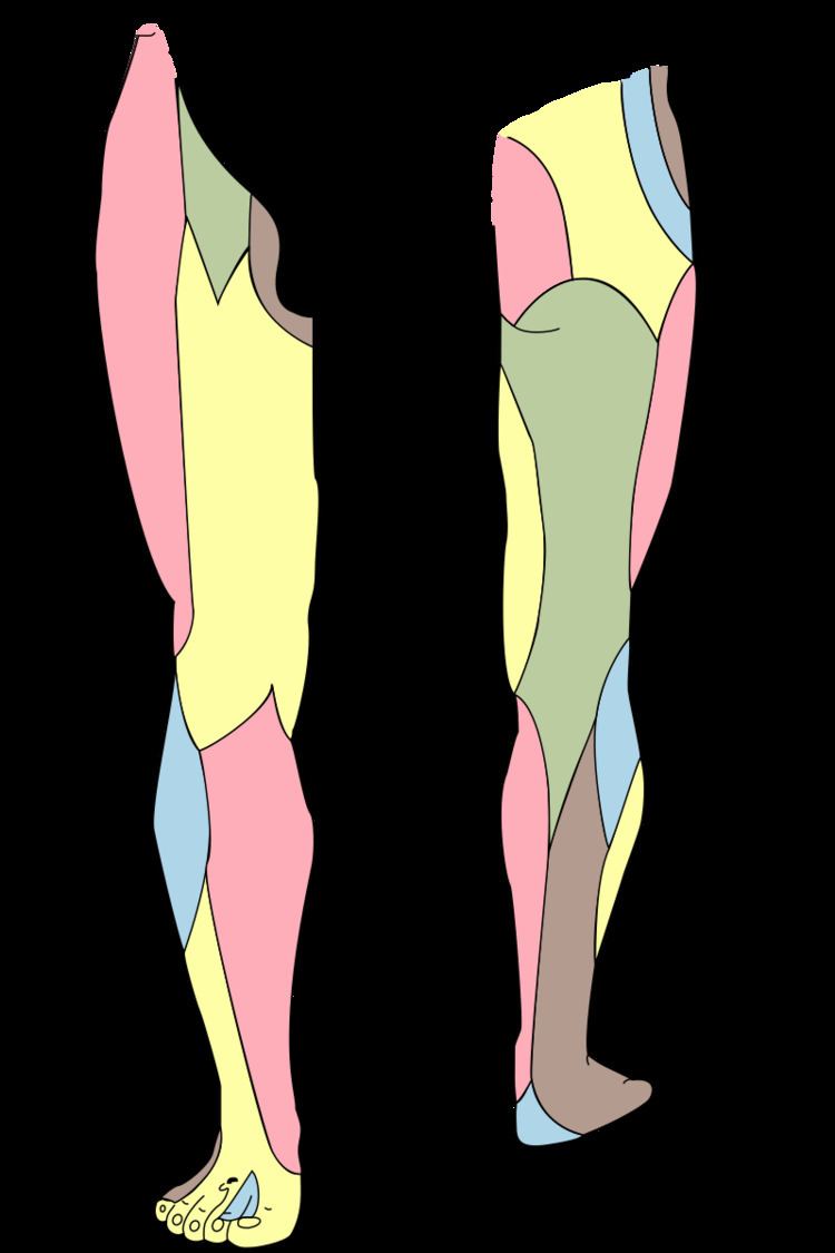
It descends obliquely along the lateral side of the popliteal fossa to the head of the fibula, close to the medial margin of the biceps femoris muscle. Where the common peroneal nerve winds round the head of the fibula, it is palpable.
It lies between the tendon of the biceps femoris and lateral head of the gastrocnemius muscle, winds around the neck of the fibula, between the peroneus longus and the bone, and divides beneath the muscle into the superficial peroneal nerve and deep peroneal nerve.
Branches
Previous to its division it gives off articular and lateral sural cutaneous nerves.
Function
The common peroneal nerve innervates the short head of the biceps femoris muscle via a motor branch that exits close to the gluteal cleft. The remainder of the peroneal-innervated muscles are innervated by its branches, the deep peroneal nerve and superficial peroneal nerve.
It provides sensory innervation to the skin over the upper third of the lateral aspect of the leg via the lateral cutaneous nerve of the calf. It gives the peroneal communicating nerve which joins the sural nerve in the midcalf.
Clinical significance
Chronic peroneal neuropathy can result from, among other conditions, bed rest of long duration, hyperflexion of the knee, peripheral neuropathy, pressure in obstetric stirrups, and conditioning in ballet dancers. The most common cause is habitual leg crossing that compresses the common peroneal nerve as it crosses around the head of the fibula. Transient trauma to the nerve can result from peroneal strike.
Damage to this nerve typically results in foot drop, where dorsiflexion of the foot is compromised and the foot drags (the toe points) during walking; and in sensory loss to the dorsal surface of the foot and portions of the anterior, lower-lateral leg. A common yoga kneeling exercise, the Vajrasana, has been linked to a variant called yoga foot drop.
Surgical procedures involving the nerve involve: