
ICD-9-CM 448.1 MedlinePlus 001441 | DiseasesDB 30744 eMedicine derm/73 | |
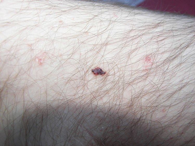 | ||
Cherry angiomas, also known as Campbell De Morgan spots or senile angiomas, are cherry red papules on the skin containing an abnormal proliferation of blood vessels. They are the most common kind of angioma. They are called Campbell de Morgan spots after the nineteenth-century British surgeon Campbell De Morgan, who first noted and described them.
Contents
The frequency of cherry angiomas increases with age.
Signs and symptoms
Cherry angiomas are made up of clusters of capillaries at the surface of the skin, forming a small round dome ("papule"), which may be flat topped. They range in colour from bright red to purple. When they first develop, they may be only a tenth of a millimeter in diameter and almost flat, appearing as small red dots. However, they then usually grow to about one or two millimeters across, and sometimes to a centimeter or more in diameter. As they grow larger, they tend to expand in thickness, and may take on the raised and rounded shape of a dome. Multiple adjoining angiomas are said to form a polypoid angioma. Because the blood vessels comprising an angioma are so close to the skin's surface, cherry angiomas may bleed profusely if they are injured.
One study found that the majority of capillaries in cherry hemangiomas are fenestrated because of staining for carbonic anhydrase activity.
Cause
Cherry angiomas appear spontaneously in many people in middle age but can also, less commonly, occur in young people. They can also occur in an aggressive eruptive manner in any age. The underlying cause for the development of cherry angiomas is not understood.
Findings suggest that cherry angioma may occur through two different mechanisms: angiogenesis (the formation of new blood vessels from pre-existing vessels), and vasculogenesis (the formation of totally new vessels, which usually occurs during embryonic and fetal development).
The first study trying to bring light to the molecular and genetic mechanisms behind cherry/senile hemangioma was published in 2010. The study found that the level of microRNA 424 is significantly reduced in senile hemangiomas compared to normal skin resulting in increased protein expression of MEK1 and cyclin E1. By inhibiting mir-424 in normal endothelial cells they could observe the same increased protein expression of MEK1 and cyclin E1 which, important for the development of senile hemangioma, induced cell proliferation of the endothelial cells. They also found that targeting MEK1 and cyclin E1 with small interfering RNA decreased the number of endothelial cells.
Chemicals and compounds that have been seen to cause cherry angiomas are mustard gas, 2-butoxyethanol, bromides, and cyclosporine.
A significant increase in the density of mast cells has been seen in cherry hemangiomas compared with normal skin.
Treatment
These lesions generally do not require treatment. If they are cosmetically unappealing or are subject to bleeding angiomas may be removed by electrocautery, a process of destroying the tissue by use of a small probe with an electric current running through it. Removal may cause scarring. More recently pulsed dye laser or Intense Pulsed Light (IPL) treatment has also been used.
Future treatment based on a locally acting inhibitor of MEK1 and Cyclin E1 could possibly be an option. A natural MEK1 inhibitor is myricetin
Prognosis
In most patients, the number and size of cherry angiomas increases with advancing age. They are harmless, having no relation to cancer at all.
Epidemiology
Cherry angiomas occur in all races, ethnic backgrounds, and sexes.