
MeSH D002557 | ||
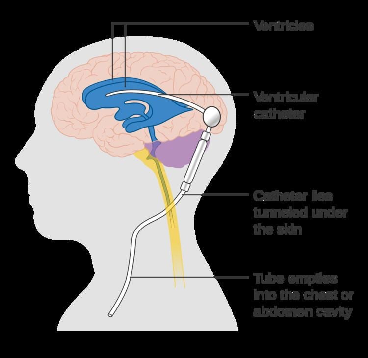 | ||
Cerebral shunts are commonly used to treat hydrocephalus, the swelling of the brain due to excess buildup of cerebrospinal fluid (CSF). If left unchecked, the cerebrospinal fluid can build up leading to an increase in intracranial pressure (ICP) which can lead to intracranial hematoma, cerebral edema, crushed brain tissue or herniation. The cerebral shunt can be used to alleviate or prevent these problems in patients who suffer from hydrocephalus or other related diseases. Shunts can come in a variety of forms but most of them consist of a valve housing connected to a catheter, the end of which is usually placed in the peritoneal cavity. The main differences between shunts are usually in the materials used to construct them, the types of valve (if any) used, and whether the valve is programmable or not.
Contents
- Shunt location
- Shunt routing
- Complications
- Infection
- Treatment of shunt infections
- Medical treatment of shunt infection
- Surgical treatment of shunt infection
- Obstruction
- Over drainage
- Chiari I malformation
- Slit ventricle syndrome
- Intraventricular hemorrhage
- Conditions requiring shunting
- Removing shunts
- References
Shunt location
The location of the shunt is determined by the neurosurgeon based on the type and location of the blockage causing hydrocephalus. All brain ventricles are candidates for shunting. The catheter is most commonly placed in the abdomen but other locations include the heart and lungs. Shunts can often be named after the route used by the neurosurgeon. The distal end of the catheter can be located in just about any tissue with enough epithelial cells to absorb the incoming CSF. Below are some common routing plans for cerebral shunts.
Shunt routing
A subgaleal shunt is usually a temporary measure used in infants who are too small or premature to tolerate other shunt types. The surgeon forms a pocket beneath the epicranial aponeurosis (the subgaleal space) and allows CSF to drain from the ventricles, creating a fluid-filled swelling on the baby's scalp. These shunts are normally converted to VP or other shunt types once the infant is big enough.
Complications
There are a number of complications associated with shunt placement. Many of these complications occur during childhood and cease once the patient has reached adulthood. Many of the complications seen in patients require immediate shunt revision (the replacement or reprogramming of the already existing shunt). The common symptoms often resemble the new onset of hydrocephalus such as headaches, nausea, vomiting, double-vision, and an alteration of consciousness. Furthermore, in the pediatric population, the shunt failure rate 2 years after implantation has been estimated to be as high as 50%.
Infection
Infection is a common complication that normally affects pediatric patients because they have not yet built up immunities to a number of different diseases. Normally, the incidence of infection decreases as the patient grows older and the body gains immunity to various infectious agents. Shunt infection is a common problem and can occur in up to 27% of patients with a shunt. Infection can lead to long term cognitive defects, neurological problems, and in some cases death. Common microbial agents for shunt infection include Staphylococcus epidermidis, Staphylococcus aureus, and Candida albicans. Further factors leading to shunt infection include shunt insertion at a young age (<6 months old) and the type of hydrocephalus being treated. There is no strong correlation between infection and shunt type. The symptoms of a shunt infection are very similar to the symptoms seen in hydrocephalus but can also include fever and elevated white blood cell counts.
Treatment of shunt infections
Treatment of a CSF shunt infection generally includes removal of the shunt and placement of a temporary ventricular reservoir until the infection is resolved. There are four main methods of treating ventriculoperitoneal (VP) shunt infections: (1) antibiotics; (2) removal of infected shunt with immediate replacement; (3) externalization of shunt with eventual replacement; (4) removal of infected shunt with external ventricular drain (EVD) placement and eventual shunt re-insertion. The last method is best with over 95% success rate.
Medical treatment of shunt infection
Initial empiric therapy for CSF shunt infection should include broad coverage that includes gram-negative aerobic bacilli including pseudomonas and gram-positive organisms including Staph aureus and coagulase negative staphylococcus, such as a combination of ceftazidime and vancomycin. Some clinicians add either parenteral or intrathecal aminoglycosides to provide enhanced pseudomonas coverage, although the efficacy of this is not clear at this time. Meropenem and aztreonam are additional options that are effective against gram-negative bacterial infections.
Surgical treatment of shunt infection
To evaluate the benefit of surgical shunt removal or externalization followed by removal, Wong et al. compared two groups: one with medical treatment alone and another with medical and surgical treatment simultaneously. 28 patients suffering from infection after ventriculoperitoneal shunt implantation over an 8-year period in their neurosurgical center were studied. 17 of these patients were treated with shunt removal or externalization followed by removal in addition to IV antibiotics while the other 11 were treated with IV antibiotics only. The group receiving both surgical shunt removal and antibiotics showed lower mortality – 19% versus 42% (p = 0.231). Despite the fact that these results are not statistically significant, Wong et al. suggest managing VP shunt infections via both surgical and medical treatment.
An analysis of 17 studies published over the past 30 years regarding children with CSF shunt infections revealed that treating with both shunt removal and antibiotics successfully treated 88% of 244 infections, while antibiotic therapy alone successfully treated the CSF shunt infection in only 33% of 230 infections.
While typical surgical methods of handling VP shunt infections involve removal and reimplantation of the shunt, different types of operations have used with success in select patients. Steinbok et al. treated a case of recurrent VP shunt infections in an eczematous patient with a ventriculosubgaleal shunt for two months till the eczema healed completely. This type of shunt allowed them to avoid the area of diseased skin that acted as the source of infection. Jones et al. have treated 4 patients with non-communicating hydrocephalus that suffered VP shunt infections with shunt removal and third ventriculostomy. These patients were cured of the infection and have not required shunt re-insertion, thus showing the effectiveness of this procedure in these types of patients.
Obstruction
Another leading cause of shunt failure is the blockage of the shunt at either the proximal or distal end. At the proximal end the shunt valve can become blocked due to the buildup of excess protein in the CSF. The extra protein will collect at the point of drainage and slowly clog the valve. The shunt can also become blocked at the distal end if the shunt is pulled out of the abdominal cavity (in the case of VP shunts), or from similar protein buildup. Other causes of blockage are overdrainage and slit ventricle syndrome.
Over drainage
Over drainage occurs when a shunt has not been adequately designed for the particular patient. Overdrainage can lead to a number of different complications some of which are highlighted below.
Usually one of two types of overdrainage can occur. First when the CSF drains too rapidly, a condition known as extra-axial fluid collection can occur. In this condition the brain collapses on itself resulting in the collection of CSF or blood around the brain. This can cause severe brain damage by compressing the brain. Furthermore, a subdural hematoma may develop. Extra-axial fluid collection can be treated in three different ways depending on the severity of the condition. Usually the shunt will be replaced or reprogrammed to release less CSF and the fluid collected around the brain will be drained. The second condition known as slit ventricle syndrome occurs when CSF slowly overdrains, over several years. More information on slit ventricle syndrome appears below.
Chiari I malformation
Recent studies have shown that over drainage of CSF due to shunting can lead to acquired Chiari I Malformation. It was previously thought that Chiari I Malformation was a result of a congenital defect but new studies have shown that overdrainage of Cysto-peritoneal shunts used to treat arachnoid cysts can lead to the development of posterior fossa overcrowding and tonsillar herniation, the latter of which is the classic definition of Chiari Malformation I. Common symptoms include major headaches, hearing loss, fatigue, muscle weakness and loss of cerebellum function.
Slit ventricle syndrome
Slit ventricle syndrome is an uncommon disorder associated with shunted patients, but results in a large number of shunt revisions. The condition usually occurs several years after shunt implantation. The most common symptoms are similar to normal shunt malfunction, but there are several key differences. First the symptoms are often cyclical and will appear and then subside several times over a lifetime. Second, the symptoms can be alleviated by lying prone. In the case of shunt malfunction neither time nor postural position will affect the symptoms.
The condition is often thought to occur during a period where overdrainage and brain growth occur simultaneously. In this case the brain fills the intraventricular space, leaving the ventricles collapsed. Furthermore, the compliance of the brain will decrease, which prevents the ventricles from enlarging, thus reducing the chance for curing the syndrome. The collapsed ventricles can also block the shunt valve, leading to obstruction. Since the effects of slit ventricle syndrome are irreversible, constant care in managing the condition is needed.
Intraventricular hemorrhage
An intraventricular hemorrhage can occur at any time during or after a shunt insertion or revision. The hemorrhage can cause an impairment in shunt function which can lead to severe neurological deficiencies. Studies have shown that intraventricular hemorrhage can occur in nearly 31% of shunt revisions.
Conditions requiring shunting
Below is a short list of known complications that can lead to hydrocephalus requiring shunting.
Removing shunts
Though there have been many cases of patients reaching "shunt independence", there is no common accord in which doctors can agree in which a patient might survive without a shunt. Another problem with shunt removal is that it is very difficult to discern when a patient might be shunt independent without very specific conditions. Overall shunt removal is a rare but not unheard of procedure.