
 | ||
A catheterization laboratory or cath lab is an examination room in a hospital or clinic with diagnostic imaging equipment used to visualize the arteries of the heart and the chambers of the heart and treat any stenosis or abnormality found.
Contents
Equipment
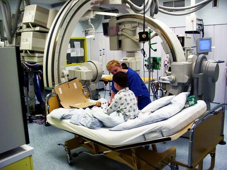
Most catheterization laboratories are "single plane" facilities, those that have a single X-ray generator source and an image intensifier. Older cath labs used cine film to record the information obtained, but since 2000, most new facilities are digital. The latest digital cath labs are biplane (have two X-ray sources) and digital, flat panel labs.
A typical 'Cath Lab' will consist of:
Staff
Cardiac catheterisation laboratories (or Cath Lab) in the UK are usually staffed by a multidisciplinary team. This includes a Medical Practitioner (normally either a Consultant Cardiologist or Radiologist), a Cardiac Physiologist, a Nurse and a Radiographer.
Medical Practitioner The Consultant Cardiologist is responsible for gaining arterial access, inserting a sheath into either the radial or femoral artery, passing a wire and catheter into the coronary artery and selectively injecting contrast media into the coronary arteries. They then interpret the images taken to ascertain where the narrowed or blocked artery has the problem. They use a variety of techniques and imaging tools to work the size of things such as balloons and stents.
Cardiac Physiologist Cardiac Physiologists usually set up what is known as a transducer to monitor pressure in the arteries. They also have a live view of the patients ECG so they can tell whether or not there is a problem being caused by the insertion of the catheter into the heart to the electrical pathways. The physiologist will also set up a temporary pacemaker if the procedure is an angioplasty or a PCI. Finally, they also set up defibrillators on to the patient for emergency use if needed.
Procedures
"Cardiac catheterization" is a general term for a group of procedures that are performed using this method, such as coronary angiography, as well as left ventricle angiography. Once the catheter is in place, it can be used to perform a number of procedures including angioplasty, PCI (percutaneous coronary intervention) angiography, balloon septostomy, and an Electrophysiology study or Catheter ablation.
A list of procedures carried out in a Cath Lab includes:
Projections
Any combination of the following views are used in visualising the Coronary Arteries:
As a general rule, cranial views are best used to demonstrate the Left Anterior Descending Artery or 'LAD', and caudal views are best used to demonstrate the Circumflex Artery or LCX.