
 | ||
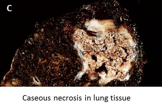
Caseous necrosis is a form of cell death in which the tissue maintains a cheese-like appearance. The dead tissue appears as a soft and white proteinaceous dead cell mass.
Contents
Causes
Frequently, caseous necrosis is encountered in the foci of tuberculosis infections. It can also be caused by syphilis and certain fungi.
A similar appearance can be associated with histoplasmosis, cryptococcosis, and coccidioidomycosis.
Appearance
In caseous necrosis no histological architecture is preserved. On microscopic examination with H&E staining, it is characterized by acellular pink areas of necrosis surrounded by a granulomatous inflammatory process.
When the hilar lymph node for instance is infected with tuberculosis and leads to caseous necrosis, its gross appearance can be a cheesy tan to white, which is why this type of necrosis is often depicted as a combination of both coagulative and liquefactive necrosis.
However, in the lung, extensive caseous necrosis with confluent cheesy tan granulomas is typical. The tissue destruction is so extensive that there are areas of cavitation (also known as cystic spaces). See Ghon's complex.