
Specialty cardiology ICD-9-CM 429.3 MeSH D006332 | ICD-10 I51.7 DiseasesDB 30769 | |
 | ||
Cardiomegaly is a medical condition in which the heart is enlarged. It is more commonly referred to as an enlarged heart. The causes of cardiomegaly may vary. Many times this condition results from high blood pressure (hypertension) or coronary artery disease. An enlarged heart may not pump blood effectively, resulting in congestive heart failure. Cardiomegaly may improve over time, but many people with an enlarged heart need lifelong treatment with medications. Having an immediate family member who has or had cardiomegaly may indicate that a person is more susceptible to getting this condition. Cardiomegaly is not a disease but rather a condition that can result from a host of other diseases such as obesity or coronary artery disease. Recent studies suggest that cardiomegaly is associated with a higher risk of sudden cardiac death (SCD).
Contents
Mechanism
Cardiomegaly is a condition affecting the cardiovascular system, specifically the heart. This condition is strongly associated with congestive heart failure. Within the heart, the working fibers of the myocardial tissue increase in size. As the heart works harder the actin and myosin filaments experience less overlap which increases the size of the myocardial fibers. If there is less overlap of the protein filaments actin and myosin within the sarcomeres of muscle fibers, they will not be able to effectively pull on one another. If the heart tissue (walls of left and right ventricle) gets too big and stretches too far, then those filaments cannot effectively pull on one another to shorten the muscle fibers, thus impacting the heart's sliding filament mechanism. If fibers cannot shorten properly, and the heart cannot contract properly, then blood cannot be effectively pumped to the lungs to be re-oxygenated and to the body to deliver oxygen to the working tissues of the body.
Signs and symptoms
For many people cardiomegaly is asymptomatic. For others, if the enlarged heart begins to affect the body's ability to pump blood effectively, then symptoms associated with congestive heart failure may arise.
Diagnosis
There are two main types of cardiomegaly:
Dilated cardiomyopathy is the most common type of cardiomegaly. In this condition, the walls of the left and/or right ventricles of the heart become thin and stretched. The result is an enlarged heart.
In the other types of cardiomegaly, the heart's large muscular left ventricle becomes abnormally thick. Hypertrophy is usually what causes left ventricular enlargement. Hypertrophic cardiomyopathy is typically an inherited condition. There are many techniques and tests used to diagnose an enlarged heart. Below is a list of tests and how they test for cardiomegaly:
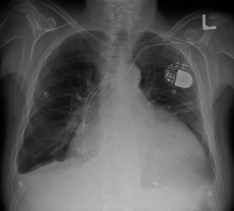
1. Chest X-Ray: X-ray images help see the condition of the lungs and heart. If the heart is enlarged on an X-ray, other tests will usually be needed to find the cause. A useful measurement on X-ray is the cardio-thoracic ratio, which is the transverse diameter of the heart, compared with that of the thoracic cage." These diameters are taken from PA chest x-rays using the widest point of the chest and measuring as far as the lung pleura, not the lateral skin margins. If the cardiac thoracic ratio is greater than 50%, pathology is suspected, assuming the x-ray has been taken correctly. The measurement was first proposed in 1919 to screen military recruits. A newer approach to using these x-rays for evaluating heart health, takes the ratio of heart area to chest area and has been called the two-dimensional cardiothoracic ratio.
2. Electrocardiogram: This test records the electrical activity of the heart through electrodes attached to the person's skin. Impulses are recorded as waves and displayed on a monitor or printed on paper. This test helps diagnose heart rhythm problems and damage to a person's heart from a heart attack.
3. Echocardiogram: This test for diagnosing and monitoring an enlarged heart uses sound waves to produce a video image of the heart. With this test, the four chambers of the heart can be evaluated.
4. Stress test: A stress test, also called an exercise stress test, provides information about how well the heart works during physical activity.
5. Cardiac computerized tomography (CT) or magnetic resonance imaging (MRI). In a cardiac CT scan, one lies on a table inside a machine called a gantry. An X-ray tube inside the machine rotates around the body and collects images of the heart and chest.
6. Blood tests: Blood tests may be ordered to check the levels of substances in the blood that may show a heart problem. Blood tests can also help rule out other conditions that may cause one's symptoms.
7. Cardiac catheterization and biopsy: In this procedure, a thin tube (catheter) is inserted in the groin and threaded through the blood vessels to the heart, where a small sample (biopsy) of the heart, if indicated, can be extracted for laboratory analysis.
Cause and prevention
The cause of cardiomegaly is not well understood and many cases of cardiomegaly are idiopathic (having no known cause). Prevention of cardiomegaly starts with detection. If a person has a family history of cardiomegaly, one should let one's doctor know so that treatments can be implemented to help prevent worsening of the condition. In addition, prevention includes avoiding certain lifestyle risk factors such as tobacco use and controlling one's high cholesterol, high blood pressure, and diabetes. Non-lifestyle risk factors include family history of cardiomegaly, coronary artery disease (CAD), congenital heart failure, Atherosclerotic disease, valvular heart disease, exposure to cardiac toxins, sleep disordered breathing (such as sleep apnea), sustained cardiac arrhythmias, abnormal electrocardiograms, and cardiomegaly on chest X-ray. Lifestyle factors which can help prevent cardiomegaly include eating a healthy diet, controlling blood pressure, exercise, medications, and not abusing alcohol and cocaine. Current research and the evidence of previous cases link the following (below) as possible causes of cardiomegaly.
The most common causes of Cardiomegaly are congenital (patients are born with the condition based on a genetic inheritance), high blood pressure which can enlarge the left ventricle causing the heart muscle to weaken over time, and coronary artery disease that creates blockages in the heart's blood supply, which can bring on a cardiac infarction (heart attack) leading to tissue death which causes other areas of the heart to work harder, increasing the heart size.
Other possible causes include:
Treatment and prognosis
Treatments for cardiomegaly include a combination of medication treatment and medical/surgical procedures. Below are some of the treatment options for individuals with cardiomegaly:
Medications
Medical devices to regulate the heartbeat
Pacemaker: Coordinates the contractions between the left and right ventricle. In people who may be at risk of serious arrhythmias, drug therapy or an implantable cardioverter-defibrillator (ICD) may be an used.
Surgical procedures
Heart valve surgery: If an enlarged heart is caused by a problem with one of the heart valves, one may have surgery to remove the valve and replace it with either an artificial valve or a tissue valve from a pig, cow or deceased human donor. If blood leaks backward through a valve (valve regurgitation), the leaky valve may be surgically repaired or replaced.
Coronary bypass surgery: If an enlarged heart is related to coronary artery disease, one may opt to have coronary artery bypass surgery.
Left ventricular assist device: (LVAD): This implantable mechanical pump helps a weak heart pump. LVADs are often implanted while a patient waits for a heart transplant or, if the patient is not a heart transplant candidate, as a long-term treatment for heart failure.
Heart transplant: If medications can't control the symptoms, a heart transplant is often a final option.
Cardiomegaly can progress and certain complications are common: