
Species Human Entrez 1000 | Human Mouse Ensembl ENSG00000170558 | |
 | ||
Aliases CDH2, CD325, CDHN, CDw325, NCAD, cadherin 2 External IDs MGI: 88355 HomoloGene: 20424 GeneCards: CDH2 | ||
N-cadherin, also known as Cadherin-2 (CDH2) or neural cadherin (NCAD) is a protein that in humans is encoded by the CDH2 gene. CDH2 has also been designated as CD325 (cluster of differentiation 325). N-cadherin is a transmembrane protein expressed in multiple tissues and functions to mediate cell–cell adhesion. In cardiac muscle, N-cadherin is an integral component in adherens junctions residing at intercalated discs, which function to mechanically and electrically couple adjacent cardiomyocytes. While mutations in CDH2 have not thus far been associated with human disease, alterations in expression and integrity of N-cadherin protein has been observed in various forms of disease, including human dilated cardiomyopathy.
Contents
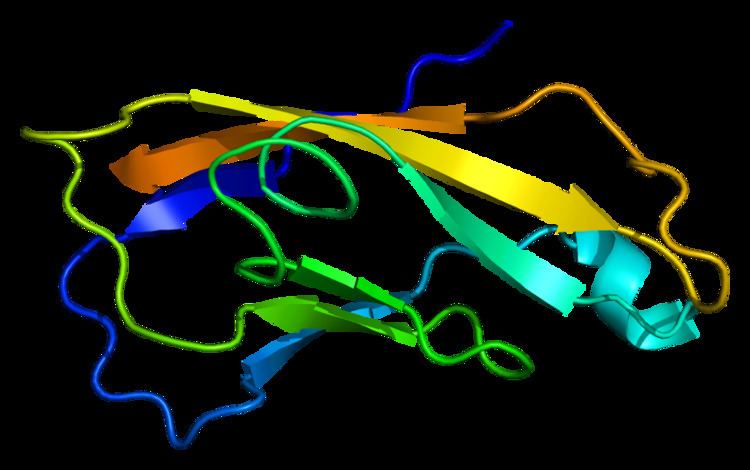
Structure
N-cadherin is a protein with molecular weight of 99.7 kDa, and 906 amino acids in length. N-cadherin a classical cadherin from the cadherin superfamily, composed of five extracellular cadherin repeats, a transmembrane region and a highly conserved cytoplasmic tail. N-cadherin, as well as other cadherins, interact with N-cadherin on an adjacent cell in an anti-parallel conformation, thus creating a linear, adhesive "zipper" between cells.
Function
N-cadherin, originally named for its role in neural tissue, plays a role in neurons and later was found to also play a role in cardiac muscle and in cancer metastasis. N-cadherin is a transmembrane, homophilic glycoprotein belonging to the calcium-dependent cell adhesion molecule family. These proteins have extracellular domains that mediate homophilic interactions between adjacent cells, and C-terminal, cytoplasmic tails that mediate binding to catenins, which in turn interact with the actin cytoskeleton.
Role in development
N-cadherin plays a role in development as a calcium dependent cell–cell adhesion glycoprotein that functions during gastrulation and is required for establishment of left-right asymmetry.
N-cadherin is widely expressed in the embryo post-implantation, showing high levels in the mesoderm with sustained expression through adulthood. N-cadherin mutation during development has the most significant effect on cell adhesion in the primitive heart; dissociated myocytes and abnormal heart tube development occur. N-cadherin plays a role in the development of the vertebrate heart at the transition of epithelial cells to trabecular and compact myocardial cell layer formation. An additional study showed that myocytes expressing a dominant negative N-cadherin mutant showed significant abnormalities in myocyte distribution and migration towards the endocardium, resulting in defects in trabecular formation within the myocardium.
Role in cardiac muscle
In cardiac muscle, N-cadherin is found at intercalated disc structures which provide end-on cell–cell connections that facilitate mechanical and electrical coupling between adjacent cardiomyocytes. Within intercalated discs are three types of junctions: adherens junctions, desmosomes and gap junctions; N-cadherin is an essential component in adherens junctions, which enables cell–cell adhesion and force transmission across the sarcolemma. N-cadherin complexed to catenins has been described as a master regulator of intercalated disc function. N-cadherin appears at cell–cell junctions prior to gap junction formation, and is critical for normal myofibrillogenesis. Expression of a mutant form of N-cadherin harboring a large deletion in the extracellular domain inhibited the function of endogenous N-cadherin in adult ventricular cardiomyocytes, and neighboring cardiomyocytes lost cell–cell contact and gap junction plaques as well.
Mouse models employing transgenesis have highlighted the function of N-cadherin in cardiac muscle. Mice with altered expression of N-cadherin and/or E-cadherin showed a dilated cardiomyopathy phenotype, likely due to malfunction of intercalated discs. In agreement with this, mice with ablation of N-cadherin in adult hearts via a cardiac-specific tamoxifen-inducible Cre N-cadherin transgene showed disrupted assembly of intercalated discs, dilated cardiomyopathy, impaired cardiac function, decreased sarcomere length, increased Z-line thickness, decreases in connexin 43, and a loss in muscular tension. Mice died within two months of transgene expression, mainly due to spontaneous ventricular tachycardia. Further analysis of N-cadherin knockout mice revealed that the arrhythmias were likely due to ion channel remodeling and aberrant Kv1.5 channel function. These animals showed a prolonged action potential duration, reduced density of inward rectifier potassium channel and decreased expression of Kv1.5, KCNE2 and cortactin combined with disrupted actin cytoskeleton at the sarcolemma.
Role in neurons
In neural cells, at certain central nervous system synapses, presynaptic to postsynaptic adhesion is mediated at least in part by N-cadherin. N-cadherins interact with catenins to play an important role in learning and memory (For full article see Cadherin-catenin complex in learning and memory).
Role in cancer metastasis
N-Cadherin is commonly found in cancer cells and provides a mechanism for transendothelial migration. When a cancer cell adheres to the endothelial cells of a blood vessel it up-regulates the src kinase pathway, which phosphorylates beta-catenins attached to both N-cadherin (this protein) and E-cadherins. This causes the intercellular connection between two adjacent endothelial cells to fail and allows the cancer cell to slip through.
Clinical significance
Mutations in CDH2 have not been conclusively linked to any human disorders. One study investigating genetic underpinnings of obsessive-compulsive disorder and Tourette disorder found that while CDH2 variants are likely not disease-causing as single entities, they may confer risk when examined as part of a panel of related cell–cell adhesion genes. Further studies in larger cohorts will be required to unequivocally determine this.
In human dilated cardiomyopathy, it was shown that N-cadherin expression was enhanced and arranged in a disarrayed fashion, suggesting that disorganization of N-cadherin protein in heart disease may be a component of remodeling.
Interactions
CDH2 has been shown to interact with: