
Specialty pulmonology ICD-9-CM 516.8 eMedicine radio/117 | ICD-10 J84.0 DiseasesDB 31684 MeSH D018549 | |
 | ||
Bronchiolitis obliterans organizing pneumonia (BOOP), also known as cryptogenic organizing pneumonia, is a form of non-infectious pneumonia; more specifically, BOOP is an inflammation of the bronchioles (bronchiolitis) and surrounding tissue in the lungs. It is often a complication of an existing chronic inflammatory disease such as rheumatoid arthritis, dermatomyositis, or it can be a side effect of certain medications such as amiodarone. BOOP was first described by Gary Epler in 1985.
Contents
Some authors have recommended the use of an alternate name, cryptogenic organizing pneumonia (COP), to reduce confusion with bronchiolitis obliterans, a distinct and unrelated disease.
The clinical features and radiological imaging resemble infectious pneumonia. However, diagnosis is suspected after there is no response to multiple antibiotics, and blood and sputum cultures are negative for organisms.
Terminology
"Organizing" refers to unresolved pneumonia (in which the alveolar exudate persists and eventually undergoes fibrosis) in which fibrous tissue forms in the alveoli. The phase of resolution and/or remodeling following bacterial infections is commonly referred to as organizing pneumonia, both clinically and pathologically.
Signs and symptoms
The classic presentation of COP is the development of nonspecific systemic (e.g., fevers, chills, night sweats, fatigue, weight loss) and respiratory (e.g. difficulty breathing, cough) symptoms in association with filling of the lung alveoli that is visible on chest x-ray. This presentation is usually so suggestive of an infection that the majority of patients with COP have been treated with at least one failed course of antibiotics by the time the true diagnosis is made.
Causes
It was identified in 1985, although its symptoms had been noted before but not recognised as a separate lung disease. The risk of BOOP is higher for people with inflammatory diseases like lupus, dermatomyositis, rheumatoid arthritis, and scleroderma.
Diagnosis
On clinical examination, crackles are common, and more rarely, patients may have clubbing (<5% of cases). Laboratory findings are nonspecific.
Almost 75% of people have symptoms for less than two months before seeking medical attention. A flu-like illness, with a cough, fever, a feeling of illness (malaise), fatigue, and weight loss heralds the onset in about 40% of patients. Doctors do not find any specific abnormalities on routine laboratory tests or on a physical examination, except for the frequent presence of crackling sounds (called rales) upon auscultation with a stethoscope by the care provider. Pulmonary function tests usually show that the amount of air the lungs can hold is below normal. The amount of oxygen in the blood is often low at rest and is even lower with exercise.
Imaging
The chest x-ray is distinctive with features that appear similar to an extensive pneumonia, with both lungs showing widespread white patches. The white patches may seem to migrate from one area of the lung to another as the disease persists or progresses. Computed tomography (CT) may be used to confirm the diagnosis. Often the findings are typical enough to allow the doctor to make a diagnosis without ordering additional tests. To confirm the diagnosis, a doctor may perform a lung biopsy using a bronchoscope. Many times, a larger specimen is needed and must be removed surgically.
Plain chest radiography shows normal lung volumes, with characteristic patchy unilateral or bilateral consolidation. Small nodular opacities occur in up to 50% of patients and large nodules in 15%. On high resolution computed tomography, airspace consolidation with air bronchograms is present in more than 90% of patients, often with a lower zone predominance A subpleural or peribronchiolar distribution is noted in up to 50% of patients. Ground glass appearance or hazy opacities associated with the consolidation are detected in most patients.
Pulmonary physiology is restrictive with a reduced diffusion capacity of the lung for carbon monoxide (DLCO). Airflow limitation is uncommon; gas exchange is usually abnormal and mild hypoxemia is common. Bronchoscopy with bronchoalveolar lavage reveals up to 40% lymphocytes, along with more subtle increases in neutrophils and eosinophils. In patients with typical clinical and radiographic features, a transbronchial biopsy that shows the pathologic pattern of organizing pneumonia and lacks features of an alternative diagnosis is adequate to make a tentative diagnosis and start therapy. On surgical lung biopsy, the histopathologic pattern is organizing pneumonia with preserved lung architecture; this pattern is not exclusive to BOOP and must be interpreted in the clinical context.
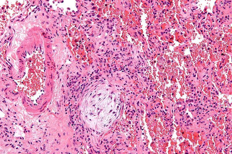
Histologically, cryptogenic organizing pneumonia is characterized by the presence of polypoid plugs of loose organizing connective tissue (Masson bodies) within alveolar ducts, alveoli, and bronchioles.
Complications
Rare cases of BOOP have induced with lobar cicatricial atelectasis.
Treatment
Most patients recover with corticosteroid therapy. A standardized approach to dosing starting at 0.75 mg/kg and weaning over 24 weeks has been shown to reduce total corticosteroid exposure without affecting outcome.
About two thirds of patients recover with corticosteroid therapy: the usual corticosteroid administered is prednisolone in Europe and prednisone in the USA; these differ by only one functional group and have the same clinical effect. The corticosteroid is initially administered in high dosage, typically 50 mg per day tapering down to zero over a six-month to one-year period. If the corticosteroid treatment is halted too quickly the disease may return. Other medications must be taken to counteract side effects of the steroid.