
 | ||
Bone tissue, or osseous tissue, is the major structural and supportive connective tissue of the body. Bone tissue forms the rigid part of the bones that make up the skeleton.
Contents
Bones are organs that are made up of bone tissue as well as bone marrow, small blood vessels, epithelium and nerves. Bone tissue refers specifically to the bone mineral matrix that forms the rigid sections of the organ, and the bone cells within it. The two types of bone tissue are cortical bone and cancellous bone. There is another kind of tissue called subchondral bone which underlies the epiphyseal cartilage at the ends of bones.
The bone cells develop new bone tissue and continual bone remodeling – maintaining the bones and the regulation of minerals in the body. Types of bone cell include osteoclasts, which break down bone tissue; osteoblasts, which build new bone tissue; osteocytes, which hold up the bone together; and lining cells, which protect the bone.
Structure
There are two types of bone tissue: cortical bone and cancellous bone: The tissues are biologically identical; the difference is in how the microstructure is arranged.
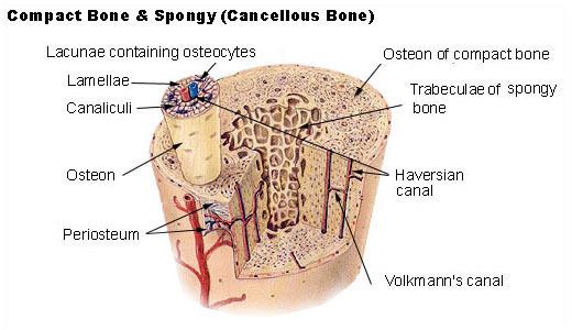
Cortical bone, synonymous with compact bone, is one of the two types of bone tissue that form bones. Cortical bone forms the extremely hard exterior of bones. Cortical bone facilitates bone's main functions: to support the whole body, protect organs, provide levers for movement, and store and release chemical elements, mainly calcium. As its name implies, cortical bone forms the cortex, or outer shell, of most bones. Cortical (compact) bone is much denser than cancellous bone. Furthermore, it is harder, stronger and stiffer than cancellous bone. Cortical bone contributes about 80% of the weight of a human skeleton. The primary anatomical and functional unit of cortical bone is the osteon.
Cancellous bone is synonymous with trabecular or spongy bone. Cancellous bone has a higher surface area to mass ratio than cortical bone because it is less dense. This makes it softer, and weaker but more flexible. The greater surface area also makes it suitable for metabolic activities such as the exchange of calcium ions. Cancellous bone is typically found at the ends of long bones, near to joints and within the interior of vertebrae. Cancellous bone is highly vascular and frequently contains red bone marrow where haematopoiesis, the production of blood cells, occurs. The primary anatomical and functional unit of cancellous bone is the trabecula. The trabeculae are aligned towards the mechanical load distribution that a bone experiences within long bones such as the femur. As far as short bones are concerned, trabecular alignment has been studied in the vertebral pedicle. The macroscopic yield strength of cancellous bone has also been investigated, using high resolution computer models.
The words cancellous and trabecular refer to the tiny lattice-shaped units (trabeculae) that form the tissue. It was first illustrated accurately in the engravings of Crisóstomo Martinez.
Bone cells
The bone cells include osteocytes, osteoblasts, osteoclasts, osteogenic cells (stem cells), and lining cells.
Osteoclasts are very large multinucleate cells that are responsible for the breakdown of bones. The breakdown of bone is very important in bone health because it allows for bone remodeling. Osteoclasts are formed by the conjoining of many different cells created from the bone marrow that travel in the circulatory system. Osteoclasts are usually found grouped in the small pits on bone surfaces called Howship's lacunae. The pits are sites of bone resorption. After latching on to the site of the bone where it is supposed to resorb it, the osteoclast releases a number of enzymes which break down the bone tissue. The final product of the resorption of the bone is calcium and phosphorus ions. This reabsorption process can sometimes take up to weeks for the osteoclast to complete. The breakdown of bones is controlled by hormones in the bloodstream which instruct the osteoclasts when and where to break down bone tissue.
Osteoblasts are bone cells that are responsible for the formation of new bone. Osteoblasts deposit a collagen matrix and release minerals that combine to make the bone mineral. Unlike the much larger osteoclasts, osteoblasts are much smaller; they only have one nucleus. Osteoblasts also group to form new bone. Osteoblasts are important because they allow the bones to be made, remodeled, and repaired. The osteoblasts come from the differentiation of osteogenic cells in the tissue that covers the outside of the bone, or the periosteum and the bone marrow. The osteoblast creates and repairs new bone by actually building around itself. First, the osteoblast puts up collagen fibers. These collagen fibers are used as a framework for the osteoblasts' work. The osteoblast then deposits calcium phosphate which is hardened by hydroxide and bicarbonate ions. The brand new bone created by the osteoblast is called osteoid. Once the osteoblast is finished working it is actually trapped inside of the bone once it hardens. When the osteoblast becomes trapped, it becomes known as an osteocyte. Other osteoblasts remain on the top of the new bone and are used to protect the underlying bone, these become known as lining cells.
Osteocytes are osteoblasts which have become trapped inside of the bone matrix. Once an osteoblast creates the new bone around itself, it is trapped and can no longer move or form bone; this is how an osteocyte is created. When the osteoblast is transformed into an osteocyte, the transformation causes the osteocyte to lose a majority of its organelles. What replaces these organelles are large quantities of microfilaments. Osteocytes develop long branches which allow them to contact each other and also contact the bone lining cells. The osteocyte lies within a small chamber called a lacuna, which is within the bone matrix. Osteocytes remain in contact with other cells in the bone through gap junctions—coupled cell processes—which pass through small channels in the bone matrix called the canaliculi. The osteocyte is still a mysterious cell—biologists still have not figured out the true function of the osteocyte. Even though the function of the osteocyte is still under investigation, there are some ideas on what they might do. One function of the osteocyte might be the remodeling of the bone through growths of new arms on the cell. It is also known that osteocytes can secrete growth factors which activate lining cells or stimulate osteoblasts. Finally, it is believed that the osteocyte might compensate for the strain on the bone due to their many arms which extend out to other osteocytes.
Lining cells come from osteoblasts which have become flattened. Bone lining cells have flat organelles so they can easily cover the bone without interfering with other cells functions. Bone lining cells are mainly in adults, some however are in the bones of children. Bone lining cells are connected to other bone lining cells through gap junctions and are able to send cell processes through canaliculi. The lining cells are relatively inactive forms of osteoblasts that cover all available surfaces of the bone. Bone lining cells are responsible for the immediate release of [calcium] in the bone if calcium in the blood is too low. Bone lining cells are also responsible for the protection of the bone from harmful chemicals which would eat away the bone. It is also thought that bone lining cells are important in the maintenance of the bone fluids.
Bone cells create molecules made from proteins which are used to communicate with other bone cells. These molecules are called growth factors and cytokines. These factors control cell division, differentiation, and survival of the cells.
Growth factors include bone morphogenetic protein (BMP) and insulin-like growth factors. BMPs are made in the bone marrow and bind to BMP receptors on the mesenchymal stem cells. Once the BMP is bound with the BMP receptor, it causes the cell to produce Cbfa 1, which is a factor that activates DNA so protein can be created. When Cbfa 1 activates genes the cells differentiate into mature osteoblasts. This is important because without the bone morphogenetic proteins the bone cells would turn into fat cells. The second type of growth factor is insulin-like growth factors. Insulin-like growth factors are created by cells such as osteoblasts, osteocytes, and bone lining cells. They are produced in the bone matrix where they gather and are released during the process of bone remodeling by osteoclasts. The main function of the insulin-like growth factors is to start bone cell replication and to help bone cells divide.
The cytokines include interleukin-1, interleukin-6, and tumor necrosis factors. A second type of cytokine is RANKL. Interleukin-1, interleukin-6, and tumor necrosis factors are made by osteoblasts, osteocytes, and bone lining cells. These cytokines cause bone marrow stem cells to differentiate, and changes in the differentiation of osteoblasts. RANKL is a cytokine that remains on the surface of osteoblasts, osteocytes, and bone lining cells. The bone cells make RANKL because of hormones and other cytokines. RANKL is a main factor of the differentiation of osteoclasts.
Formation
Bone tissue is a mineralized connective tissue. It is formed by cells, called osteoblasts, that deposit a matrix of Type-I collagen and also release calcium, magnesium, and phosphate ions that ultimately combine chemically within the collagenous matrix to form a crystalline mineral, known as bone mineral, in the form of carbonated hydroxyapatite. The combination of hard mineral and flexible collagen makes bone harder and stronger than cartilage without being brittle. Cortical bone consists of a repeating structure called an osteon or haversian system, which is the primary anatomical and functional unit. Each osteon has concentric lamellae (layers) of mineralized matrix, which are deposited around a central canal, known as the haversian canal, each containing blood and nerve supply.
Function
Bone tissue performs numerous functions including: Directly:
Indirectly:
Clinical significance
In functioning to maintain pH balance, the loss of calcium carbonate can cause a bone-softening condition known as osteomalacia due to the inability to construct new bone. Certain conditions such as kidney disease cause a build-up of hydrogen ions in the blood which will initiate the release of calcium carbonate. Like osteoporosis, osteomalacia carries the higher risk of fractures.
Osteopenia is a condition of decreased bone density that is caused by a number of factors including ageing. It can progress to more serious osteoporosis.
In postmenopausal osteoporosis, cancellous bone is more severely affected than cortical bone.
Bone tumors can affect the bone tissue and these can be benign or malignant.