
 | ||
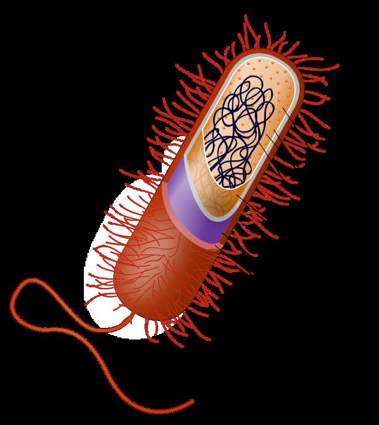
The cell capsule is a very large structure of some prokaryotic cells, such as bacterial cells. It is a polysaccharide layer that lies outside the cell envelope of bacteria, and is thus deemed part of the outer envelope of a bacterial cell. It is a well-organized layer, not easily washed off, and it can be the cause of various diseases.
Contents
The capsule—which can be found in both Gram-negative bacteria and Gram-positive bacteria—should not be confused with the second lipid membrane (or bacterial outer membrane), which contains lipopolysaccharides and lipoproteins and is found only in Gram-negative bacteria. When the amorphous viscid secretion (that makes up the capsule) diffuses into the surrounding medium and remains as a loose undemarcated secretion, it is known as slime layer.
Composition
It usually consists of polysaccharides, but can be composed of other materials (e.g., polypeptide(D-glutamic acid) in B. anthracis),also peptidoglycan and muramic acid found in E.coli bacterial capsule. Because most capsules are so tightly packed, they are difficult to stain using standard stains because most stains cannot adhere to the capsule. For examination under the microscope, the bacteria and their background are stained darker than the capsule, which doesn't stain. When viewed, bacterial cells as well as the surface they are on, are stained dark, while the capsule remains pale or colorless and appears as a ring, or halo, around the cell.
Function
The capsule is considered a virulence factor because it enhances the ability of bacteria to cause disease (e.g. prevents phagocytosis). The capsule can protect cells from engulfment by eukaryotic cells, such as macrophages. A capsule-specific antibody may be required for phagocytosis to occur. Capsules also contain water which protects the bacteria against desiccation. They also exclude bacterial viruses and most hydrophobic toxic materials such as detergents. Immunity to one capsule type does not result in immunity to the other types. Capsules also help cells adhere to surfaces.
Diversity
The capsule is found most commonly among Gram-negative bacteria:
However, some Gram-positive bacteria may also have a capsule:
The yeast Cryptococcus neoformans, though not a bacterium, has a similar capsule.
Capsules too small to be seen with an ordinary microscope, such as the M protein of Streptococcus pyogenes, are called microcapsules.
Mnemonic
A common mnemonic used to remember some encapsulated pathogens is:
"Even Some Super Killers Have Pretty Nice Big Capsules"
Escherichia coli, Streptococcus pneumoniae, Salmonella, Klebsiella pneumoniae, Haemophilus influenzae, Pseudomonas aeruginosa, Neisseria meningitidis, Bacteroides fragilis, and the yeast Cryptococcus neoformans.
Demonstration of capsule
- India ink staining: the capsule appears as a clear halo around the bacterium as the ink can't penetrate the capsule.
- Maneval's capsule stain: the capsule appears as a clear halo between the pink-stained bacterium and the bluish-grey stained background. The background stain is the acidic stain Congo red (which changes color to bluish-grey due to the pH), and the pink stain is acid fuschin.
- Serological methods: Capsular material is antigenic and can be demonstrated by mixing it with a specific anticapsular serum. When examined under the microscope, the capsule appears 'swollen' due to an increase in its refractivity. This phenomenon is the basis of Quellung reaction.
Use in vaccination
Vaccination using capsular material is effective against some organisms (e.g., H. influenzae type b, S. pneumoniae, and N. meningitidis). However, polysaccharides are not highly antigenic, especially in children, so many capsular vaccines contain polysaccharides conjugated with protein carriers, such as the tetanus toxoid or diphtheria toxoid. This stimulates a much more robust immune response.