
 | ||
Similar Bumper fracture, Burst fracture, Calcaneal fracture | ||
An ankle fracture is a break of the ankle bones. It is typically diagnosed by X-ray. Treatment is with splinting, casting or surgery. In children ankle fractures occur in about 1 per 1000 per year.
Contents
Signs and symptoms
Symptoms of an ankle fracture can be similar to those of ankle sprains (pain), though typically they are often more severe by comparison. It is exceedingly rare for the ankle joint to dislocate in the presence of ligamentous injury alone. However, in the setting of an ankle fracture the talus can become unstable and subluxate or dislocate. Patients may complain of ecchymosis (bruising), or there may be an abnormal position, abnormal motion, or lack of motion.
Evaluation
On clinical examination, it is important to evaluate the exact location of the pain, the range of motion and the condition of the nerves and vessels. It is important to palpate the calf bone (fibula) because there may be an associated fracture proximally (Maisonneuve fracture), and to palpate the sole of the foot to look for a Jones fracture at the base of fifth metatarsal (avulsion fracture).
Evaluation of ankle injuries for fracture is done with the Ottawa ankle rules, a set of rules that were developed to minimize unnecessary X-rays. There are three x-ray views in a complete ankle series: anteroposterior, lateral, and oblique (or "mortise view"). The mortise view an anteroposterior x-ray taken with the ankle internally rotated until the lateral malleolus is on the same horizontal plane as the medial malleolus, and a line drawn through both malleoli would be parallel to the tabletop, resulting in a position where there normally is no superimposition of tibia and fibula on each other. It usually requires 10 to 20 degrees of internal rotation.
X-ray
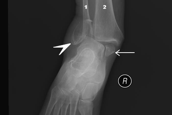
On X-rays, there can be a fracture of the medial malleolus, the lateral malleolus, or of the anterior/posterior margin of the distal tibia. The posterior margin (known as the posterior malleolus) is much more frequently injured than the anterior aspect of the distal tibia. If both the lateral and medial malleoli are broken, this is called a bimalleolar fracture (some of them are called Pott's fractures). If the posterior malleolus is also fractured, this is called a trimalleolar fracture.
Classification
There are several classification schemes for ankle fractures:
Treatment
Treatment of ankle fractures is dictated by the stability of the ankle joint. Certain fractures patterns are deemed stable, and may be treated similar to ankle sprains. All other types require surgery, most often an open reduction and internal fixation (ORIF), which is usually performed with permanently implanted metal hardware that holds the bones in place while the natural healing process occurs. A cast or splint will be required to immobilize the ankle following surgery.
In children recovery may be faster with an ankle brace rather than a full cast in those with otherwise stable fractures.
Epidemiology
In children ankle fractures occur in about 1 per 1000 per year.