
 | ||
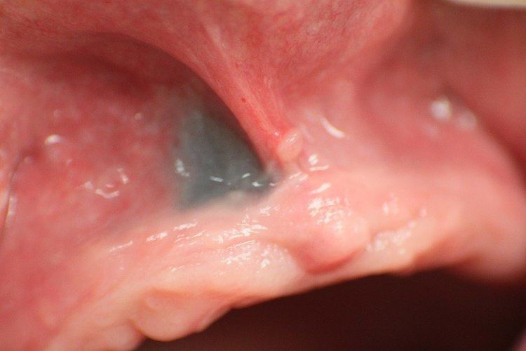
Amalgam tattoo (also termed localized argyrosis, or focal argyrosis) is a grey, blue or black area of discoloration on the mucous membranes of the mouth, typically on the gums of the lower jaw. It is an iatrogenic lesion, caused by entry of dental amalgam into the soft tissues. It is common, painless, and benign, but it can be mistaken for melanoma.
Contents
Signs and symptoms
Amalgam tattoo usually occurs on the mandibular gingiva, often in an area in which a apicoectomy ("root-end filling") with amalgam was carried out. After the gingiva, the alveolar mucosa and the buccal mucosa are the next most common sites, although any mucosal site in the mouth is possible. It is painless, and appears as a blue-black or grey discolored macule on the surface of the mucosa. The borders of the tattoo are variable, and may be well defined, irregular or diffuse.
Causes
Amalgam tattoo is caused by implantation of amalgam into the tissues. It may occur in several ways:
Over time, the amalgam particles embedded in the soft tissues corrode. Macrophages take up the exogenous particles, and the silver in amalgam leads to staining of collagen fibers.
A similar appearance can be caused by implantation of graphite (e.g. from pencil leads), and is sometimes termed a graphite tattoo, although this is less common than tattooing with amalgam.
Diagnosis
The diagnosis is clinical. Amalgam tattoo can be distinguished from other causes of localized oral pigmentation because it does not change significantly in size or color, although it may appear to slowly enlarge for several months after the initial implantation of the metal particles. Some amalgam tattoos appear radio-opaque on radiographs (i.e. they show up on x-rays), although in many cases amalgam tattoos have no radiographic features since the responsible particle(s) of amalgam are very small even though clinically the area of discolored mucosa is much larger.
If necessary, the diagnosis can be confirmed histologically by excisional biopsy, which excludes nevi and melanomas. If a biopsy is taken, the histopathologic appearance is:
Prevention
Theoretically, routine use of a dental dam during dental procedures which involve amalgam should reduce the risk of amalgam tattoo.
Treatment
No treatment is required since the lesion is entirely benign. Some suggest that amalgam tattoos are best surgically excised so as to ensure the lesion does not represent a melanoma. Other say that excision should only be carried out if there is any doubt over the diagnosis, and that amalgam tattoos are managed by simple reassurance about the nature of the lesion. For example, if radio-opaque particles are demonstrated on the x-ray, biopsy is unnecessary.
Epidemiology
Amalgam tattoo is found in up to 1% of people in the general population. It is the most common cause of solitary or focal pigmentation of the oral mucosa.