
Symbol ADK InterPro IPR000850 SCOP 1ake | Pfam PF00406 PROSITE PDOC00104 SUPERFAMILY 1ake | |
 | ||
Adenylate kinase (EC 2.7.4.3) (also known as ADK or myokinase) is a phosphotransferase enzyme that catalyzes the interconversion of adenine nucleotides, and plays an important role in cellular energy homeostasis.
Contents
Substrate and products
The reaction catalyzed is:
ATP + AMP ⇔ 2 ADP
The equilibrium constant varies with condition, but it is close to 1. Thus, ΔGo for this reaction is close to zero. In muscle from a variety of species of vertebrates and invertebrates, the concentration of ATP is typically 7-10 times that of ADP, and usually greater than 100 times that of AMP. The rate of oxidative phosphorylation is controlled by the availability of ADP. Thus, the mitochondrion attempts to keep ATP levels high due to the combined action of adenylate kinase and the controls on oxidative phosphorylation.
ADK isozymes
This is an essential reaction for many processes in living cells. Two ADK isozymes have been identified in mammalian cells. These specifically bind AMP and favor binding to ATP over other nucleotide triphosphates (AK1 is cytosolic and AK2 is located in the mitochondria). A third ADK has been identified in bovine heart and human cells. This is a mitochondrial GTP:AMP phosphotransferase, also specific for the phosphorylation of AMP, but can only use GTP or ITP as a substrate. ADK has also been identified in different bacterial species and in yeast. Two further enzymes are known to be related to the ADK family, i.e. yeast uridine monophosphokinase and slime mold UMP-CMP kinase. Within the ADK family there are several conserved regions, including the ATP-binding domains. One of the most conserved areas includes an Arg residue, whose modification inactivates the enzyme, together with an Asp that resides in the catalytic cleft of the enzyme and participates in a salt bridge.
Subfamilies
Isozymes
Human genes encoding proteins with adenylate kinase include:
Mechanism
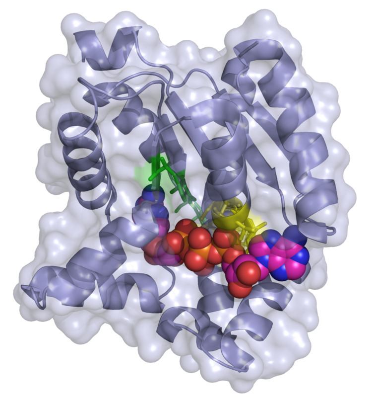
In Escherichia coli, the crystal structure of ADK was analyzed in a 2005 study. The crystal structure revealed that ADK was complexed with diadenosine pentaphosphate (AP5A), Mg2+, and 4 coordinated water molecules. ATP adenine and ribose moieties are loosely bound to ADK. The phosphates in ATP are strongly bound to surrounding residues. Mg2+, coordination waters, and surrounding charged residues maintain the geometry and distances of the AMP α-phosphate and ATP β- and γ-phosphates. And, this is sufficient to support an associative reaction mechanism for phosphoryl transfer. ADK catalyzes the transfer of a phosphoryl group from ATP to AMP by nucleophilic attack on the γ-phosphate of ATP.
Structure
Flexibility and plasticity allow proteins to bind to ligands, form oligomers, aggregate, and perform mechanical work. Large conformational changes in proteins play an important role in cellular signaling. Adenylate Kinase is a signal transducing protein; thus, the balance between conformations regulates protein activity. ADK has a locally unfolded state that becomes depopulated upon binding.
A 2007 study by Whitford et al. shows the conformations of ADK when binding with ATP or AMP. The study shows that there are three relevant conformations or structures of ADK—CORE, Open, and Closed. In ADK, there are two small domains called the LID and NMP. ATP binds in the pocket formed by the LID and CORE domains. AMP binds in the pocket formed by the NMP and CORE domains.
The study also reported findings that show that localized regions of a protein unfold during conformational transitions. This mechanism reduces the strain and enhances catalytic efficiency. Local unfolding is the result of competing strain energies in the protein. The interconversion between inactive (open) and active (closed) conformations is rate limiting for catalysis.
Metabolic monitoring
ADK uses AMP metabolic signals produced or downregulated during exercise, stress response, food consumption, hormone changes. ADK relays deliver AMP signals to metabolic sensors. It facilitates decoding of cellular information by catalyzing nucleotide exchange in the intimate “sensing zone” of metabolic sensors.
Through a chain of sequential reactions, ADK facilitates transfer and utilization of γ- and β-phosphoryls in the ATP molecule.
ADK shuttle
The energy of two high-energy phosphoryls, γ- and β-phosphoryls in the ATP molecule, is made available by the ADK present in mitochondrial and myofibrillar compartments. ATP and AMP are transferred between ATP-production and ATP-consumption sites that involve multiple, sequential phosphotransfer relays. This results in a flux wave propagation along groups of ADK molecules. This ligand conduction mechanism facilitates metabolic flux without apparent changes in metabolite concentrations.
ADK reads the cellular energy state, generates, tunes, and communicates AMP signals to metabolic sensors. In this way, ADK is able to convey information about the overall energy balance. AMP-sensors inhibit ATP consumption and promote ATP production.
Nucleoside diphosphate kinase deficiency
Nucleoside diphosphate (NDP) kinase catalyzes in vivo ATP-dependent synthesis of ribo- and deoxyribonucleoside triphosphates. In mutated Escherichia coli that had a disrupted nucleoside diphosphate kinase, adenylate kinase performed dual enzymatic functions. ADK complements nucleoside diphosphate kinase deficiency.
Hemolytic anemia
Adenylate kinase deficiency in the erythrocyte is associated with hemolytic anemia. This is a rare hereditary erythroenzymopathy that, in some cases, is associated with mental retardation and psychomotor impairment. At least two patients have exhibited neonatal icterus and splenomegaly and required blood transfusions due to this deficiency. In another patient, an abnormal fragment with homozygous and heterozygous A-->G substitutions at codon 164 caused severe erythrocyte ADK deficiency. Two siblings had erythrocyte ADK deficiency, but one did not have evidence of hemolysis.
AK1 and post-ischemic coronary reflow
Knock out of AK1 disrupts the synchrony between inorganic phosphate and turnover at ATP-consuming sites and ATP synthesis sites. This reduces the energetic signal communication in the post-ischemic heart and precipitates inadequate coronary reflow flowing ischemia-reperfusion.
ADK2 deficiency
Adenylate Kinase 2 (AK2) deficiency in humans causes hematopoietic defects associated with sensorineural deafness. Recticular dysgenesis is an autosomal recessive form of human combined immunodeficiency. It is also characterized by an impaired lymphoid maturation and early differentiation arrest in the myeloid lineage. AK2 deficiency results in absent or a large decrease in the expression of proteins. AK2 is specifically expressed in the stria vascularis of the inner ear which indicates why individuals with an AK2 deficiency will have sensorineural deafness.
Structural adaptations
AK1 genetic ablation decreases tolerance to metabolic stress. AK1 deficiency induces fiber-type specific variation in groups of transcripts in glycolysis and mitochondrial metabolism. This supports muscle energy metabolism.
Plastidial ADK deficiency in Arabidopsis thaliana
Enhanced growth and elevated photosynthetic amino acid is associated with plastidial adenylate kinase deficiency in Arabidopsis thaliana.