
 | ||
In ocular physiology, adaptation is the ability of the eye to adjust to various levels of darkness and light.
Contents
Efficiency
The human eye can function from very dark to very bright levels of light; its sensing capabilities reach across nine orders of magnitude. This means that the brightest and the darkest light signal that the eye can sense are a factor of roughly 1,000,000,000 apart. However, in any given moment of time, the eye can only sense a contrast ratio of 1,000. What enables the wider reach is that the eye adapts its definition of what is black.
The eye takes approximately 20–30 minutes to fully adapt from bright sunlight to complete darkness and become 10,000 to 1,000,000 times more sensitive than at full daylight. In this process, the eye's perception of color changes as well (this is called the Purkinje effect). However, it takes approximately five minutes for the eye to adapt from darkness to bright sunlight. This is due to cones obtaining more sensitivity when first entering the dark for the first five minutes but the rods take over after five or more minutes.
Dark adaptation is far quicker and deeper in young people than the elderly.
Ambient light response
A minor mechanism of adaptation is the pupillary light reflex, adjusting the amount of light that reaches the retina.
In response to varying ambient light levels, rods and cones of eye function both in isolation and in tandem to adjust the visual system. Changes in the sensitivity of rods and cones in the eye are the major contributors to dark adaptation.
Above a certain luminance level (about 0.03 cd/m2), the cone mechanism is involved in mediating vision; photopic vision. Below this level, the rod mechanism comes into play providing scotopic (night) vision. The range where two mechanisms are working together is called the mesopic range, as there is not an abrupt transition between the two mechanism. This adaptation forms the basis of the Duplicity Theory.
Dark adaptation
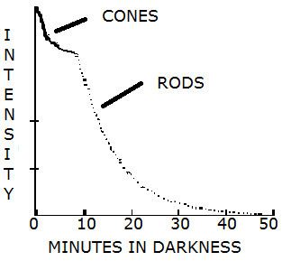
Rhodopsin, a biological pigment in the photoreceptors of the retina, immediately photobleaches in response to light. Rods are more sensitive to light and so take longer to fully adapt to the change in light. Rods, whose photopigments regenerate more slowly, do not reach their maximum sensitivity for about half an hour. Cones take approximately 9–10 minutes to adapt to the dark. Sensitivity to light is modulated by changes in intracellular calcium ions and cyclic guanosine monophosphate.
The sensitivity of the rod pathway improves considerably within 5–10 minutes in the dark. Color testing has been used to determine the time at which rod mechanism takes over; when the rod mechanism takes over colored spots appear colorless as only cone pathways encode color.
Four factors affect dark adaptation:
Inhibition
Inhibition by neurons also affects activation in synapses. Together with the bleaching of a rod or cone pigment, merging of signals on ganglion cells are inhibited, reducing convergence. Alpha adaptation, i.e., rapid sensitivity fluctuations, is powered by nerve control. The merging of signals by virtue of the diffuse ganglion cells, as well as horizontal and amacrine cells, allow a cumulative effect. Thus that area of stimulation is inversely proportional to intensity of light, a strong stimulus of 100 rods equivalent to a weak stimulus of 1,000 rods.
In sufficiently bright light, convergence is low, but during dark adaptation, convergence of rod signals boost. This is not due to structural changes, but by a possible shutdown of inhibition that stops convergence of messages in bright light. If only one eye is open, the closed eye must adapt separately upon reopening to match the already adapted eye.
Measuring Dark Adaptation
Ophthalmologists sometimes measure patients’ dark adaptation using an instrument known as a dark adaptometer. Currently, there is one commercially available dark adaptometer, called the AdaptDx. It works by measuring a patient’s Rod Intercept (RI) time. RI is the number of minutes it takes for the eye to adapt from bright light to darkness. This RI number provides a clear and objective measurement of retinal function with 90% sensitivity and specificity. Basically, an RI of less than 6.5 minutes indicates a healthy dark adaptation function. However, an RI higher than 6.5 indicates impaired dark adaptation.
Using Dark Adaptation Measurement to Diagnose Disease
Numerous clinical studies have shown that dark adaptation function is dramatically impaired from the earliest stages of AMD, retinitis pigmentosa (RP), and other retinal diseases, with increasing impairment as the diseases progress. AMD is a chronic, progressive disease that causes a part of your retina, called the macula, to slowly deteriorate as you get older. It is also the leading cause of vision loss among people age 50 and older. It is characterized by a breakdown of the RPE/Bruch’s membrane complex in the retina, leading to an accumulation of cholesterol deposits in the macula. Eventually, these deposits become clinically-visible drusen that affect photoreceptor health, causing inflammation and a predisposition to choroidal neovascularization (CNV). During the AMD disease course, the RPE/Bruch’s function continues to deteriorate, hampering nutrient and oxygen transport to the rod and cone photoreceptors. As a side effect of this process, the photoreceptors exhibit impaired dark adaptation because they require these nutrients for replenishment of photopigments and clearance of opsin to regain scotopic sensitivity after light exposure.
Measurement of a patient’s dark adaptation function is essentially a bioassay of the health of their Bruch’s membrane. As such, research has shown that, with the AdaptDx, doctors can detect subclinical AMD at least three years earlier than it is clinically evident.
Light adaptation
With light adaptation, the eye has to quickly adapt to the background illumination to be able to distinguish objects in this background. The process for light adaptation occurs over a period of five minutes.
The photochemical reaction is:
Rhodopsin ⇌ retinal + opsinIncrement threshold
Using increment threshold experiments, light adaptation can be measured clinically. In an increment threshold experiment, a test stimulus is presented on a background of a certain luminance, the stimulus is increased until the detection threshold is reached against the background. A monophasic or biphasic threshold versus intensity TVI curve is obtained through this method for both cones and rods.
When the threshold curve for a single system (i.e., just cones or just rods) is taken in isolation it can been seen to possesses four sections:
Insufficiency
Insufficiency of adaptation most commonly presents as insufficient adaptation to dark environment, called night blindness or nyctalopia. The opposite problem, known as hemeralopia, that is, inability to see clearly in bright light, is much rarer.
The fovea is blind to dim light (due to its cone-only array) and the rods are more sensitive, so a dim star on a moonless night must be viewed from the side, so it stimulates the rods. This is not due to pupil width since an artificial fixed-width pupil gives the same results.