
 | ||
Acoustic Radiation Force Impulse (ARFI) Imaging is a type of ultrasound elastography used in medicine, particularly for the diagnosis and monitoring of cancers. ARFI imaging uses acoustic radiation force to generate images of the mechanical properties of soft tissue.
Contents
How it works
Acoustic radiation force is a phenomenon associated with the propagation of acoustic waves in attenuating media. Attenuation includes both scattering and absorption of the acoustic wave. Attenuation is a frequency dependent phenomenon, and in soft tissues it is dominated by absorption. With increasing acoustic frequencies, the tissue does not respond fast enough to the transitions between positive and negative pressures, thus its motion becomes out of phase with the acoustic wave, and energy is deposited into the tissue. This energy results in a momentum transfer in the direction of wave propagation and tissue heating. The momentum transfer generates a force that causes displacement of the tissue, and the time scale of this response is much slower than that of the ultrasonic wave propagation. This displacement (typically a few micrometers), which is typically detected by computing the correlation of ultrasonic RF signal, can be used to derive additional information about the tissue beyond what is normally provided in an ultrasonic image. The magnitude, location, spatial extent, and duration of acoustic radiation force can be controlled to interrogate the mechanical properties of the tissue.
Clinical applications
Clinical applications of ARFI imaging include:
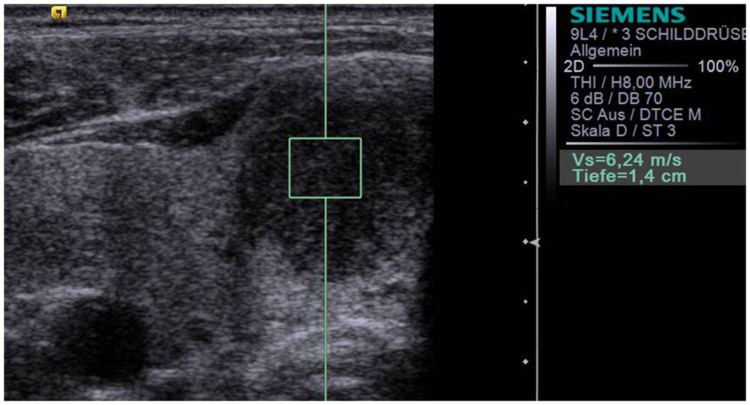
Liver fibrosis quantification
The speed at which shear waves propagate in tissue can be used to quantify the shear modulus of the tissue. Acoustic radiation force-based imaging modalities, including SWEI, are being studied to non-invasively characterize the liver without the need for liver biopsy.
Liver Fibrosis Quantification efforts at Duke University
Acoustic Radiation Force Impulse Imaging (ARFI): a New Technique to Assess Liver Elasticity
Yoneda et al. also recently compared ARFI shear wave imaging as implemented on the Siemens Acuson S2000 with transient elastography using the FibroScan system (EchoSens, Paris, France) in the context of evaluating patients with Non-alcoholic Fatty Liver Disease (NAFLD).
Breast mass imaging
Focused acoustic waves that propagate through tissue are absorbed and generate radiation force. Acoustic radiation force results from a transfer of momentum from an acoustic wave to tissue in the direction of wave propagation, arising from absorption and scattering of the wave. The tissue displacement response to radiation force excitation occurs on a slower time-scale than ultrasonic wave propagation, thus, conventional ultrasonic methods can be used to monitor the tissue response to radiation force. For breast imaging, experiments have been conducted to measure the potential for ARFI images to provide adjunctive information to matched ultrasonic images in order to enhance clinical confidence in diagnosis of breast masses. Patients scheduled for core biopsy of breast masses are recruited and matched B-mode and ARFI images of masses were obtained. The images were then correlated with biopsy result.
Colorectal tumor imaging/staging
Colorectal cancer is the second leading cause of cancer death in the United States (after lung cancer), and the third most common cancer overall in men (after prostate and lung cancer) and women (after breast and lung cancer). Once identified, the treatment approach for rectal cancer is dictated by the stage of the tumor (T-stage) and local lymph nodes (N-stage). There are currently no imaging methods that provide reliable N-staging accuracy of colorectal cancers. While endorectal ultrasound (EUS) is the standard for staging the degree of wall invasion of rectal cancers (T-stage), its accuracy is poor in the critical determination between uT2 and uT3, with 10-35% of uT2 tumors being overstaged. The consequences for the patient are dramatic. A stage uT3 rectal carcinoma is treated by radical surgical resection, and is typically treated with neoadjuvant chemotherapy and radiation. Neoadjuvant treatment damages adjacent healthy tissues, and can impair the healing process following surgery, leading to increased complications of infection, bleeding, decreased colonic mobility, and incontinence. Whereas stage uT2, N- and lower tumors generally receive a transanal local excision and are not treated neoadjuvantly, eliminating these additional risks to the patient.
The objective of ARFI Imaging development is to develop and evaluate the ability of ARFI imaging techniques to image layered tissue structures as found in the gastrointestinal tract, and to guide treatment decisions through improved preoperative tumor and lymph node staging.
Prostate imaging
Prostate cancer is the most common cancer and the second leading cause of cancer death in American men. According to the American Cancer Society (ACS), an estimated 218,890 new cases of prostate cancer will be found and 27,050 men will die of this disease in 2007. Early diagnosis is essential for better treatment and increasing survival rate. Although the current screening techniques, antigen (PSA) blood testing and digital rectal examination (DRE) are considered sensitive enough for cancer screening, follow-up biopsies have significant shortcomings. Without a good imaging technique to target the needle biopsy in the prostate gland, only about 25% of tests are positive for cancer in more than 1 million prostate biopsies performed each year; the false negative rates range from 25-45% based on the first time biopsy.
Although imaging techniques are essential for cancer diagnosis, imaging the structures and lesions within prostates has been a challenging task. The goal of ARFI imaging is to optimize and evaluate ARFI techniques for prostate imaging, which will potentially provide a more accurate, low cost and patient friendly imaging guidance for targeting prostate biopsies.