
Specialty gastroenterology MeSH D014077 | ICD-10 K03.2 | |
 | ||
Acid erosion, also known as dental erosion, is a type of tooth wear. It is defined as the irreversible loss of tooth structure due to chemical dissolution by acids not of bacterial origin. Dental erosion is the most common chronic disease of children ages 5–17, although it is only relatively recently that it has been recognised as a dental health problem. There is generally widespread ignorance of the damaging effects of acid erosion; this is particularly the case with erosion due to fruit juices, because they tend to be seen as healthy. Erosion is found initially in the enamel and, if unchecked, may proceed to the underlying dentin.
Contents
- Extrinsic acidic sources
- Intrinsic acidic sources
- Behaviour
- Diagnosis
- Diagnosis based on optical properties
- Prevention and management
- References
The most common cause of erosion is by acidic foods and drinks. In general, foods and drinks with a pH below 5.0–5.7 have been known to trigger dental erosion effects. Numerous clinical and laboratory reports link erosion to excessive consumption of drinks. Those thought to pose a risk are soft drinks, some alcohol and fruit drinks, fruit juices such as orange juice (which contain citric acid) and carbonated drinks such as colas (in which the carbonic acid is not the cause of erosion, but citric and phosphoric acid). Additionally, wine has been shown to erode teeth, with the pH of wine as low as 3.0–3.8. Other possible sources of erosive acids are from exposure to chlorinated swimming pool water, and regurgitation of gastric acids.
Extrinsic acidic sources
Acidic drinks and foods lower the pH level of the mouth so consuming them causes the teeth to demineralise. Drinks low in pH levels that cause dental erosion include fruit juices, sports drinks, wine, beer and carbonated drinks . Orange and apple juices are common culprits among fruit juices. Carbonated drinks such as colas, lemonades are also very acidic, as are fruit-flavoured drinks and dilutables. Frequency rather than total intake of acidic juices is seen as the greater factor in dental erosion; infants using feeding bottles containing fruit juices (especially when used as a comforter) are therefore at greater risk of acid erosion.
Saliva acts as a buffer, regulating the pH when acidic drinks are ingested. Drinks vary in their resistance to the buffering effect of saliva. Studies show that fruit juices are the most resistant to saliva's buffering effect, followed by, in order: fruit-based carbonated drinks and flavoured mineral waters, non-fruit-based carbonated drinks, sparkling mineral waters; Mineral water being the least resistant. Because of this, fruit juices in particular, may prolong the drop in pH levels.
A number of medications such as vitamin C, aspirin and some iron preparations are acidic and may contribute towards acid erosion.
Intrinsic acidic sources
Dental erosion can occur by non-extrinsic factors too. Intrinsic dental erosion is known as perimolysis, whereby gastric acid from the stomach comes into contact with the teeth. People with diseases such as anorexia nervosa, bulimia, and gastroesophageal reflux disease (GERD) often suffer from this. GERD is quite common and an average of 7% of adults experience reflux daily. The main cause of GERD is increased acid production by the stomach. This is not exclusive to adults, as GERD and other gastrointestinal disorders may cause dental erosions in children. Rumination also may cause acid erosion.
Behaviour
Acid erosion often coexists with abrasion and attrition. Abrasion is most often caused by brushing teeth too hard.
Frothing or swishing acidic drinks around the mouth increases the risk of acid erosion.
Diagnosis
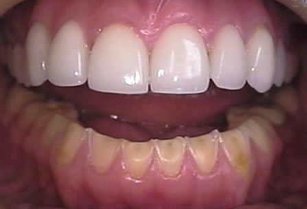
There are many signs of dental erosion, including changes in appearance and sensitivity. One of the physical changes can be the color of teeth. There are two different colors teeth may turn if dental erosion is occurring, the first being a change of color that usually happens on the cutting edge of the central incisors. This causes the cutting edge of the tooth to become transparent. A second sign is if the tooth has a yellowish tint. This occurs because the white enamel has eroded away to reveal the yellowish dentin. A change in shape of the teeth is also a sign of dental erosion. Teeth will begin to appear with a broad rounded concavity, and the gaps between teeth will become larger. There can be evidence of wear on surfaces of teeth not expected to be in contact with one another. If dental erosion occurs in children, a loss of enamel surface characteristics can occur. Amalgam restorations in the mouth may be clean and non-tarnished. Fillings may also appear to be rising out of the tooth, the appearance being caused when the tooth is eroded away leaving only the filling. The teeth may form divots on the chewing surfaces when dental erosion is occurring. This mainly happens on the first, second, and third molars. One of the most severe signs of dental erosion is cracking, where teeth begin to crack off and become coarse. Other signs include pain when eating hot, cold, or sweet foods. This pain is due to the enamel having been eroded away, exposing the sensitive dentin.
Diagnosis based on optical properties
Based on the optical changes induced in eroded tissue by the lesions, in 2015 Koshoji et al. also demonstrated in a novel method that using laser speckle images (LSI) it is possible to acquire information on the microstructure of the enamel and detect minimal changes, such as early non-carious lesions. To produce the erosion, the samples were divided into four groups and immersed in 30 ml of a cola-based beverage (pH approximately 2.5) at room temperature. A representative image of the samples under white and laser illumination shows that although there are visible stains in the left portion of each sample due the dye from the cola beverage, structural changes are difficult to assess with the naked eye.
To differentiate the sound and eroded tissues, contrast analysis was performed of the speckle patterns in the images. Since this analysis is, in its essence, the ratio of the standard deviation and average intensity, the LASCA map of the lesion is generally higher than in sound tissue. This phenomenon is demonstrated in the LASCA maps which show the greater prevalence of dark blue on the right side, indicating sound tissue, and lower prevalence on the left side, indicating eroded tissue. The contrast ratio of the LASCA maps demonstrates that laser speckle images are sensitive to even small changes in the microstructure of the surface.
Erosion is highly prevalent in people of all ages. However, an objective diagnostic procedure is still needed, thus the study of the laser speckle imaging for tooth enamel may provide the first low cost objective diagnostic method for this disease. The analysis of laser speckle imaging in the spatial domain is a powerful diagnostic technique that provides information on the surface microstructure of tooth enamel after an acid etching procedure using patterns and LASCA maps. In an erosion model, these patterns are associated with mineral loss from the enamel. This method has proven sensitive to 10 minutes of acid etching on tooth enamel, which is a lesion so incipient that is not likely to be detected in clinical practice even by a trained dentist, besides it is also sensitive to the erosion progression.
Prevention and management
Preventive and management strategies include the following: