
 | ||
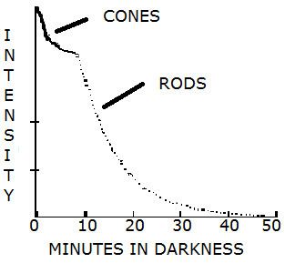
Night vision, or scotopic vision, is the ability to see under low light conditions. In humans, rod cells are exclusively responsible for night vision as cone cells are only able to function at higher illumination levels. Night vision is of a much poorer quality than day vision because it is limited by a reduced resolution and therefore provides the ability to only discriminate between shades of black and white. In order for humans to transition from day to night vision they must undergo a dark adaptation period in which each eye adjusts from a high luminescence setting to a low luminescence setting. This adaptation period is different for both rod and cone cells and results from the regeneration of photopigments to restore retinal sensitivity. Cone cells are able to regain maximum retinal sensitivity in 9–10 minutes of darkness whereas rods require 30–45 minutes to do so. There exist many ways in which humans can accelerate the dark adaptation period and therefore gain full vision in low luminescence settings in a shorter period of time.
Contents
Advantages of night vision
Many animals such as cats possess high-resolution night vision, allowing them to discriminate objects with high frequencies in low illumination settings. The tapetum lucidum is a reflective structure that is responsible for this superior night vision as it mirrors light back through the retina exposing the photoreceptor cells to an increased amount of light. Most animals which possess a tapetum lucidum are nocturnal most likely because upon reflection of light back through the retina the initial images become blurred. Humans, like their primate relatives, do not possess a tapetum lucidum and therefore were predisposed to be a diurnal species.
Despite the fact that the resolution of human day vision is far superior to that of night vision, human night vision provides many advantages. Like many predatory animals humans can use their night vision to prey upon and ambush other animals without their awareness. Furthermore, in the event of an emergency situation occurring at night humans can increase their chances of survival if they are able to perceive their surroundings and get to safety. Both of these benefits can be used to explain why humans did not completely lose the ability to see in the dark from their nocturnal ancestors.
Dark adaptation
After moving from a high luminescence setting to a low luminescence setting each eye must adjust before being they are able to achieve maximal retinal sensitivity. Upon exposure to light photopigments in both rod and cone photoreceptor cells undergo a structural change. It is through this chemical reaction known as light adaptation that light energy is converted to electrical impulses sent to the brain. During light adaptation the photopigments are decomposed thereby reducing retinal sensitivity in low luminescence settings. This is known as photoreceptor bleaching and is proportional to the quantum catch of the pigment. The regeneration of the photopigments occurs during dark adaptation albeit at markedly different rates. Rod cells are much slower to adapt to the dark and it is believed to take days for these cells to reach full dark adaptation.
Rhodopsin bleaching
The photopigment found in rod cells is called rhodopsin. Upon the absorption of light rod cells becomes bleached causing rhodopsin to lose all of its colour and become transparent. During dark adaptation the rod cells regenerate and the rhodopsin regains its purple pigmentation rendering them capable of capturing light.
Vitamin A
Vitamin A is necessary for proper functioning of the human eye. The photopigment rhodopsin found in human rod cells is composed of retinal, a form of vitamin A, bound to an opsin protein. Upon the absorption of light rhodopsin was decomposed into retinal and opsin through bleaching. Retinal could then have one of two fates: it could recombine with opsin to reform rhodopsin or it could be converted into free retinol. The American scientist George Wald was the first to recognize that the visual system expends vitamin A and is dependent upon diet for its replacement. Vitamin A serves many functions in the human body outside of healthy vision. It is vital in maintaining a healthy immune system as well as promoting normal growth and development. The average adult male and female should consume 900 and 700 micrograms of vitamin A per day, respectively. Consumption above 3000 micrograms per day is referred to as vitamin A toxicity and is usually caused by accidental ingestion of supplements.
Sources of vitamin A
Vitamin A is present in both animal and plant sources as retinoids and carotenoids, respectively. Retinoids can be used immediately by the body upon absorption into the cardiovascular system; however, plant-based carotenoids must be converted to retinol prior to utilization by the body. The highest animal-based sources of vitamin A are liver, dairy products, and fish. Fruits and vegetables containing high amounts of carotenoids are dark green, yellow, orange, and red in colour.
Evolutionary context
Vitamin A-based opsin proteins have been used for sensing light in organisms for most of evolutionary history beginning approximately 3 billion years ago. This feature has been passed from unicellular to multicellular organisms including Homo sapiens. This vitamin was most likely chosen by evolution for sensing light because retinal causes a shift in photoreceptor absorbance to the visible light range. This shift in absorbance is especially important for life on Earth because it generally matches the peak irradiance of sunlight on its surface. A second reason why retinal evolved to be vital for human vision is because it undergoes a large conformational change when exposed to light. This conformational change is believed to make it easier for the photoreceptor protein to distinguish between its silent and activated state thus better controlling visual phototransduction.
Experimental evidence
Various studies have been conducted testing the effective of vitamin A supplementation on dark adaptation. In a study by Cideciyan et al. the length of dark adaptation was measured in a patient with systemic vitamin A deficiency (VAD) before and after vitamin A supplementation. The dark adaptation function was measured prior to supplementation, 1 day post-treatment, and 75 days post-treatment. It was observed that after merely one day of vitamin A supplementation the recovery kinetics of dark adaptation were significantly accelerated after photoreceptor bleaching. Dark adaptation was further accelerated following 75 days of treatment. A subsequent study by Kemp et al. studied dark adaptation in subjects with primary biliary cirrhosis and Crohn’s disease, both of which had vitamin A deficiency. Within 8 days of oral supplementation of vitamin A both patients had their visual function restored to normal. Furthermore, adaptation kinetics significantly improved in both subjects following supplementation.
Nyctalopia
Nyctalopia (night blindness) is a condition in which one cannot see in low luminescence conditions. Night blindness can be caused by a number of factors the most common of which being vitamin A deficiency. If detected early enough nyctalopia can be reversed and visual function can be regained; however; prolonged vitamin A deficiency can lead to permanent visual loss if left untreated. Night blindness is especially prominent in developing countries due to malnutrition and therefore a lack of vitamin A in the diet. In developed countries night blindness has historically been uncommon due to adequate food availability; however, the incidence is expected to increase as obesity becomes more common. Increased obesity rates correspond to an increased number of bariatric surgeries, causing malabsorption of vitamin A in the human body.
Cones vs. rods
The human eye contains two types of photoreceptors, rods and cones, which can be easily distinguished by their structure. Cone photoreceptors are conical in shape and contain cone opsins as their visual pigments. There exist three types of cone photoreceptors, each being maximally sensitive to a specific wavelength of light depending on the structure of their opsin photopigment. The various cone cells are maximally sensitive to either short wavelengths (blue light), medium wavelengths (green light), or long wavelengths (red light). Rod photoreceptors only contain one type of photopigment, rhodopsin, which has a peak sensitivity at a wavelength of approximately 530 nanometers which corresponds to blue-green light. The distribution of photoreceptor cells across the surface of the retina has important consequences for vision. Cone photoreceptors are concentrated in a depression in the center of the retina known as the fovea centralis and decrease in number towards the periphery of the retina. Conversely, rod photoreceptors are present at high density throughout the most of the retina with a sharp decline in the fovea. Perception in high luminescence settings is dominated by cones despite the fact that they are greatly outnumbered by rods (approximately 4.5 million to 91 million).
Red lights and lenses for dark adaptation
As a result of rod cells having a peak sensitivity at a wavelength of 530 nanometers they cannot perceive all colours on the visual spectrum. Because rod cells are insensitive to long wavelengths, the use of red lights and red lens glasses has become a common practise for accelerating dark adaptation. In order for dark adaptation to be significantly accelerated an individual should ideally begin this practise 30 minutes prior to entering a low luminescence setting. This practise will allow an individual to maintain their photopic (day) vision whilst preparing for scotopic vision. The insensitivity to red light will prevent the rod cells from further becoming bleached and allow for the rhodopsin photopigment to recharge back to its active conformation. Once an individual enters a dark setting most of their rod cells will already be accommodated to the dark and be able to transmit visual signals to the brain without an accommodation period. The concept of red lenses for dark adaptation is based upon experimentation by Antoine Béclère and his early work with radiology. In 1916, the scientist Wilhelm Trendelenburg invented the first pair of red adaptation goggles for radiologists to adapt their eyes to view screens during fluoroscopic procedures.
Evolutionary context
Although many aspects the human visual system remain uncertain, the theory of the evolution of rod and cone photopigments is agreed upon by most scientists. It is believed that the earliest visual pigments were those of cone photoreceptors, with rod opsin proteins evolving later. Following the evolution of mammals from their reptilian ancestors approximately 275 million years ago there was a nocturnal phase in which complex colour vision was lost. Being that these pro-mammals were nocturnal they increased their sensitivity in low luminescence settings and reduced their photopic system from tetrachromatic to dichromatic. The shift to a nocturnal lifestyle would demand more rod photoreceptors to absorb the blue light emitted by the moon during the night. It can be extrapolated that the high ratio of rods to cones present in modern human eyes was retained even after the shift from nocturnal back to diurnal. It is believed that the emergence of trichromacy in primates occurred approximately 55 million years ago when the surface temperature of the planet began to rise. The primates were diurnal rather than nocturnal in nature and therefore required a more precise photopic visual system. A third cone photopigment was necessary to cover the entire visual spectrum enabling primates to better discriminate between fruits and detect those of the highest nutritional value.
Real world application
Aviators
Aviators commonly wear red lensed glasses or goggles prior to taking off in the dark to ensure that they are able to see outside of the aircraft. Furthermore, throughout flight the cock pit is illuminated with dim red lights. This lighting is to ensure that the pilot is able to read instruments and maps while maintaining scotopic vision for looking outside.
Submarines
Oftentimes submarines are “rigged for red,” meaning that the boat is going to be surfacing or coming to periscope depth at night. During such times illumination within certain compartments is switched to red light to allow the eyes of the lookouts and officers to adjust to the darkness prior to looking outside of the boat. Additionally, compartments on a submarine may be illuminated with red light in order to simulate night conditions for the crew.
Anthocyanins
Anthocyanins make up the majority of the 4000 known flavonoid phytochemicals. This group of approximately 600 bioactive antioxidants carries the strongest physiological effects of any plant compound. These chemicals are also the most visible of the flavonoid phytochemicals because they provide bright blue, red, or purple pigmentation to many plant species. Anthocyanins also serve to protect the photosynthetic tissues from the direct rays of the sun. In addition, the antioxidant, anti-inflammatory, and vasoprotective properties of anthocyanins allow them to demonstrate diverse health effects. In humans, anthocyanins are effective for a variety of health conditions including neurological damage, atherosclerosis, diabetes, as well as visual impairment. Anthocyanins frequently interact with other phytochemicals to potentiate biological effects; therefore, contributions from individual biomolecules remains difficult to decipher. As a result of anthocyanins providing bright colouration to flowers, the plants containing these phytochemicals are naturally successful in attracting pollinators such as birds and bees. The fruits and vegetables produced by such plants are also brightly pigmented attracting animals to eat them and disperse the seeds. Due to this natural mechanism anthocyanin-containing plants are widely abundant in most areas of the world. The high abundance and distribution of anthocyanin-containing plants make it a natural food source for many animals. Through fossil evidence it is known that these compounds were eaten in high amounts by primitive hominins.
Food sources
Brightly coloured fruits and vegetables are rich in anthocyanins. This makes sense intuitively because anthocyanins offer pigmentation to plants. Blackberries are the most anthocyanin-rich foods, containing 89-211 milligrams per 100 grams. Other foods that are rich in this phytochemical include red onions, blueberries, bilberries, red cabbage, and eggplant. The ingestion of any of these food sources will yield a variety of phytochemicals in addition to anthocyanins because they naturally exist together. The daily intake of anthocyanins is estimated to be approximately 200 milligrams in the average adult; however, this value can reach several grams per day if an individual is consuming flavonoid supplements.
Effect on dark adaptation
Anthocyanins accelerate dark adaptation in humans by enhancing the regeneration of the rod photopigment, rhodopsin. Anthocyanins accomplish this by binding directly to opsin upon the degradation of rhodopsin to its individual constituents by light. Once bound to opsin, the anthocyanin changes its structure thereby accelerating its access to the retinal binding pocket. By having a diet rich in anthocyanins an individual is able to generate rhodopsin in shorter periods of time because of the increased affinity of opsin to retinal. Through this mechanism an individual is able to accelerate dark adaptation and achieve night vision in a shorter period of time.
Experimental evidence
In a double-blind, placebo-controlled study conducted by Nakaishi et al. a powdered anthocyanin concentrate derived from black currants was provided to a number of participants. Participants received one of three doses of anthocyanins to measure if the result occurred in a dose-dependent manner. The period of dark adaptation was measured prior to and two hours following supplementation in all participants. Results from this experiment indicate that anthocyanins significantly accelerated dark adaptation at merely one dose level compared to the placebo. Observing the data as a whole Nakaishi et al. concluded that anthocyanins effectively reduced the dark adaptation period in a dose-dependent manner.
Contradictory evidence
Despite the fact that many scientists believe anthocyanins to be beneficial in accelerating dark adaptation in humans, a study conducted by Kalt et al. in 2014 showed blueberry anthocyanins have no effect. In this study two double-blind, placebo-controlled studies were conducted to examine dark adaptation following the intake of blueberry products. In neither study did the blueberry anthocyanin intake effect the length of dark adaptation. From these results Kalt et al. concluded that blueberry anthocyanins provide no significant difference to the dark adaptation component of human vision.
Real world application
During World Wars I and II British Air Force aviators were known to consume extensive amounts of bilberry jam. The aviators consumed this anthocyanin-rich food due to its many visual benefits, included accelerated dark adaptation, which would be valuable for night bombing missions.