
Origin Ribs 5-12 | Dorlands/Elsevier m_22/12549865 | |
 | ||
Insertion Iliac crest, Pubic tubercle, Linea alba Nerve Thoracoabdominal nerves (T7-11) and Subcostal nerve (T12) Latin Musculus obliquus externus abdominis | ||
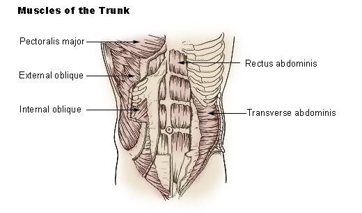
The external oblique muscle (of the abdomen) (also external abdominal oblique muscle) is the largest and the most superficial (outermost) of the three flat muscles of the lateral anterior abdomen.
Contents
Structure
The external oblique is situated on the lateral and anterior parts of the abdomen. It is broad, thin, and irregularly quadrilateral, its muscular portion occupying the side, its aponeurosis the anterior wall of the abdomen. In most humans (especially females), the oblique is not visible, due to subcutaneous fat deposits and the small size of the muscle.
It arises from eight fleshy digitations, each from the external surfaces and inferior borders of the fifth to twelfth ribs. These digitations are arranged in an oblique line which runs inferiorly and anteriorly, with the upper digitations being attached close to the cartilages of the corresponding ribs, the lowest to the apex of the cartilage of the last rib, the intermediate ones to the ribs at some distance from their cartilages.
The five superior serrations increase in size from above downward, and are received between corresponding processes of the serratus anterior muscle; the three lower ones diminish in size from above downward and receive between them corresponding processes from the latissimus dorsi. From these attachments the fleshy fibers proceed in various directions.
Those from the lowest ribs pass nearly vertically downward, and are inserted into the anterior half of the outer lip of the iliac crest; the middle and upper fibers, directed downward (inferiorly) and forward (anteriorly), become aponeurotic at approximately the midclavicular line. This aponeurosis formed from fibres from either side of the external oblique decussates at the linea alba.
The aponeurosis of the external oblique muscle forms the inguinal ligament. The muscle also contributes to the inguinal canal.
Just deep to the external oblique is the internal oblique muscle. These muscles are in the deepest layer of the abdominal wall.
Nerve supply
The external oblique muscle is supplied by ventral branches of the lower six thoracoabdominal nerves and the subcostal nerve on each side.
Blood supply
The cranial portion of the muscle is supplied by the lower intercostal arteries, whereas the caudal portion is supplied by a branches of either the deep circumflex iliac artery or the iliolumbar artery.
Function
The external oblique functions to pull the chest downwards and compress the abdominal cavity, which increases the intra-abdominal pressure as in a valsalva maneuver. It also has limited actions in both flexion and rotation of the vertebral column. One side of the obliques contracting can create lateral flexion.
Oblique strain
The oblique strain is a common baseball injury, particularly in pitchers. In both batters and pitchers it can affect the contralateral (leading) side external oblique, or the trailing internal oblique.