
Precursor mesoderm Vein trabecular vein | Artery trabecular artery Dorlands
/Elsevier n_09/12576042 | |
 | ||
Latin noduli lymphoidei splenici | ||
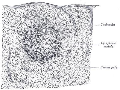
White pulp is a histological region of the spleen. The altered coat of the arterioles, consisting of adenoid tissue, presents here and there thickenings of a spheroidal shape, the white pulp (Malpighian bodies of the spleen, splenic lymphoid nodules).
These bodies vary in size from about 0.25 mm. to 1 mm. in diameter.
They are merely local expansions or hyperplasia of the adenoid tissue, of which the external coat of the smaller arteries of the spleen is formed.
They are most frequently found surrounding the arteriole, which thus seems to tunnel them, but occasionally they grow from one side of the vessel only, and present the appearance of a sessile bud growing from the arterial wall.
There are several parts of white pulp with distinct functions: