
 | ||
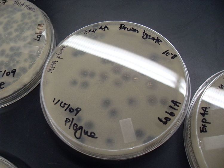
A viral plaque is a visible structure formed within a cell culture, such as bacterial cultures within some nutrient medium (e.g. agar). The bacteriophage viruses replicate and spread, thus generating regions of cell destructions known as plaques.
Counting the number of plaques can be used as a method of virus quantification. These plaques can sometimes be detected visually using colony counters, in much the same way as bacterial colonies are counted; however, they are not always visible to the naked eye, and sometimes can only be seen through a microscope, or using techniques such as staining (e.g. neutral red for eukaryotes or giemsa for bacteria) or immunofluorescence. Special computer systems have been designed with the ability to scan samples in batches.
The appearance of the plaque depends on the host strain, virus and the conditions. Highly virulent or lytic strains give clear plaques while strains that only kill a fraction of their hosts (due to partial resistance/lysogeny) or only reduce the rate of cell growth give turbid plaques. Some partially lysogenic phages give bull's-eye plaques with spots or rings of growth in the middle of clear regions of complete lysis.