
MeSH D020775 | ||
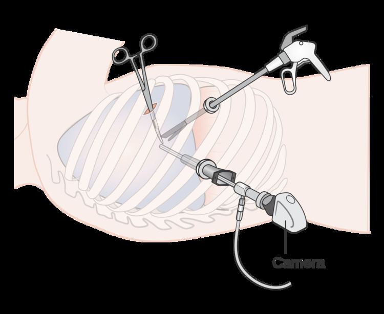 | ||
Video-assisted thoracoscopic surgery (VATS) is a type of thoracic surgery performed using a small video camera that is introduced into the patient's chest via a scope. The surgeon is able to view the instruments that are being used along with the anatomy on which the surgeon is operating. The camera and instruments are inserted through separate holes in the chest wall also known as "ports". These small ports are advantageous because the chance for infection and wound dehiscence are drastically reduced. This allows for a faster recovery by the patient and a greater chance for the wound to heal.
Traditionally, thoracic surgery performed for diagnosis or treatment of chest conditions has required access to the chest through thoracotomy or sternotomy incisions. Sternotomy requires the use of a sternal saw to divide the sternum and requires spreading of the divided portions of the sternum with a sternal retractor to allow for visualization of the thoracic structures, passage of instruments into the chest, and removal of specimens. Thoracotomy, as most commonly performed, requires division of one or more major muscles of the chest wall including the latissimus dorsi, pectoralis or serratus muscles, along with spreading of the ribs with a rib spreader. Because the joints of the ribs with the vertebral bodies have only limited flexibility, the use of a rib spreader usually results in rib fractures in the process of rendering the interspace between the ribs wide enough to perform diagnostic or therapeutic maneuvers. Because of this, thoracic surgeons generally intentionally remove a section of one or more ribs in an effort to prevent splintered rib fractures associated with the use of a rib spreader. Sternotomy and thoracotomy have been proven over decades to provide highly effective means of access to thoracic structures and in general are tolerated by patients. However, both incisions have the potential for causing significant pain that may last for extended periods and both result in bony fractures that require a minimum of six weeks to heal during which time patients must refrain from heavy lifting or strenuous activity. The great advantage of VATS over sternotomy or thoracotomy is avoidance of muscle division and bone fractures that allows for diminished duration and intensity of pain and a shorter time to return to full activity.
VATS came into widespread use beginning in the early 1990s. Operations that traditionally were carried out with thoracotomy or sternotomy that today can be performed with VATS include: biopsy for diagnosis of pulmonary, pleural or mediastinal pathology; decortication for empyema; pleurodesis for recurrent pleural effusions or spontaneous pneumothorax; surgical stapler assisted wedge resection of lung masses; resection of mediastinal or pleural masses; thoracic sympathectomy for hyperhidrosis; operations for diaphragmatic hernias or paralysis; esophageal resection or resection of esophageal masses or diverticula; and VATS lobectomy/mediastinal lymphadenectomy for lung cancer.
The instrumentation for VATS includes the use of a camera-linked 5 mm or 10 mm fiber-optic scope, with or without a 30-degree angle of visualization, and either conventional thoracic instruments or laparoscopic instruments. Unlike with laparoscopy, carbon dioxide insufflation is not generally required with VATS due to the inherent vault-like shape of the thoracic cavity. However, lung deflation on the side of the chest where VATS is being performed is a must to be able to visualize and pass instruments into the thorax; this is usually effected with a double-lumen endo-tracheal tube that allows for single lung ventilation or a bronchial blocker delivered via a standard single-lumen endotracheal tube.
Similarly to laparoscopy, VATS has enjoyed widespread use for technically straightforward operations such as pulmonary decortication, pleurodesis, and lung or pleural biopsies, while more technically demanding operations such as esophageal operations, mediastinal mass resections, or pulmonary lobectomy for early stage lung cancer, have been slower to catch on and have tended to remain confined to selected centers. It is expected that advanced VATS techniques will continue to grow in numbers spurred by patient demand and greater surgeon comfort with the techniques.