
Nerve femoral nerve | ||
 | ||
Insertion Patella via the Quadriceps tendon and Tibial tuberosity via the Patellar ligament Artery lateral circumflex femoral artery Actions Extends and stabilizes knee | ||
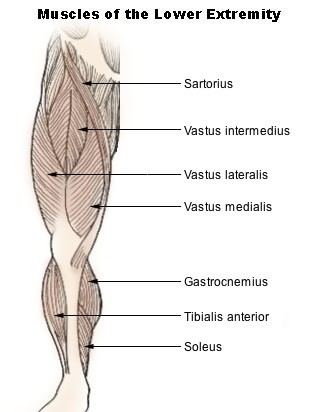
The Vastus lateralis (/ˈvæstəsˌlætəˈreɪlᵻs/ or /ˈvæstəsˌlætəˈrælᵻs/), also called the ''vastus externus'' is the largest and most powerful part of the quadriceps femoris, a muscle in the thigh. It arises from a series of flat, broad tendons attached to the femur, and attaches to the outer border of the patella. It ultimately joins with the other muscles that make up the quadriceps in the quadriceps tendon, which travels over the knee to connect to the tibia.
Structure
The vastus lateralis arises from the several areas of the femur, including the upper part of the intertrochanteric line; the lower, anterior borders of the greater trochanter, to the outer border of the gluteal tuberosity, and the upper half of the outer border of the linea aspera. These form an aponeurosis, a broad flat tendon which covers the upper three-quarters of the muscle. From the inner surface of the aponeurosis, many muscle fibres originate. Some additional fibres arise from the tendon of the gluteus maximus muscle, and from the septum between the vastus lateralis and short head of the biceps femoris.
The fibers form a large fleshy mass, attached to a second strong aponeurosis, placed on the deep surface of the lower part of the muscle. This lower aponeurosis becomes contracted and thickened into a flat tendon that attaches to the outer border of the patella, and subsequently joins with the quadriceps femoris tendon, expanding the capsule of the knee-joint.