
DiseasesDB 33482 MeSH D001327 | MedlinePlus 000821 | |
 | ||
In type II hypersensitivity (also tissue-specific, or cytotoxic hypersensitivity) the antibodies produced by the immune response bind to antigens on the patient's own cell surfaces. The antigens recognized in this way may either be intrinsic ("self" antigen, innately part of the patient's cells) or extrinsic (adsorbed onto the cells during exposure to some foreign antigen, possibly as part of infection with a pathogen). These cells are recognized by macrophages or dendritic cells, which act as antigen-presenting cells. This causes a B cell response, wherein antibodies are produced against the foreign antigen.
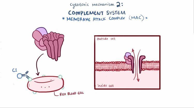
An example of type II hypersensitivity is the ABO blood incompatibility where the red blood cells have different antigens, causing them to be recognized as different; B cell proliferation will take place and antibodies to the foreign blood type are produced. IgG and IgM antibodies bind to these antigens to form complexes that activate the classical pathway of complement activation to eliminate cells presenting foreign antigens. That is, mediators of acute inflammation are generated at the site and membrane attack complexes cause cell lysis and death. The reaction takes hours to a day.
Type II reactions can affect healthy cells. Examples include red blood cells in autoimmune hemolytic anemia and acetylcholine receptors in myasthenia gravis.
Another example of type II hypersensitivity reaction is Goodpasture's syndrome where the basement membrane (containing collagen type IV) in the lung and kidney is attacked by one's own antibodies.
Another form of type II hypersensitivity is called antibody-dependent cell-mediated cytotoxicity (ADCC). Here, cells exhibiting the foreign antigen are tagged with antibodies (IgG or IgM). These tagged cells are then recognised by natural killer cells (NK) and macrophages (recognised via IgG bound (via the Fc region) to the effector cell surface receptor, CD16 (FcγRIII)), which in turn kill these tagged cells.